Copyright
©2014 Baishideng Publishing Group Inc.
World J Clin Cases. Jul 16, 2014; 2(7): 289-292
Published online Jul 16, 2014. doi: 10.12998/wjcc.v2.i7.289
Published online Jul 16, 2014. doi: 10.12998/wjcc.v2.i7.289
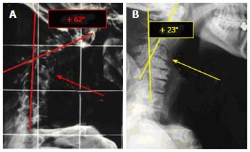
Figure 1 X-ray pictures.
A: Standard X-ray demonstrating a severe cervical kyphosis (preop. Angle of Jackson +62°); B: A cervical spine X-ray on the bed with a pillow under the shoulders showing a good reduction of kyphosis (+23° according to Jackson) due to the motor unit C4-C5 mobility.
Figure 2 The preoperative computed tomography.
A: Scan control (preop. Angle of Jackson +62°); B: Showing a good reduction of kyphosis (postop angle of Jackson +19°).
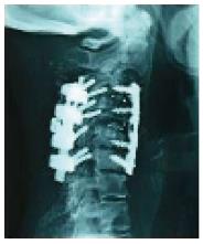
Figure 3 A postoperative X-ray control after 6 mo showing a good anterior and posterolateral arthrodesis.
- Citation: Landi A, Marotta N, Mancarella C, Dugoni DE, Tarantino R, Delfini R. 360° fusion for realignment of high grade cervical kyphosis by one step surgery: Case report. World J Clin Cases 2014; 2(7): 289-292
- URL: https://www.wjgnet.com/2307-8960/full/v2/i7/289.htm
- DOI: https://dx.doi.org/10.12998/wjcc.v2.i7.289


