Copyright
©2014 Baishideng Publishing Group Inc.
World J Clin Cases. Oct 16, 2014; 2(10): 534-540
Published online Oct 16, 2014. doi: 10.12998/wjcc.v2.i10.534
Published online Oct 16, 2014. doi: 10.12998/wjcc.v2.i10.534
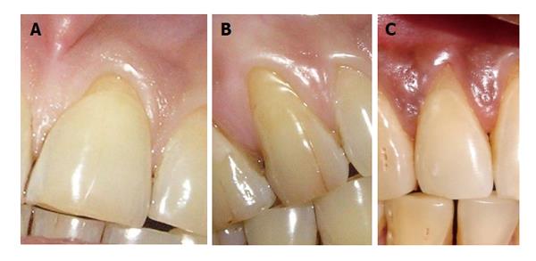
Figure 1 Miller’s class-I gingival recession.
A: Miller’s class-I gingival recession with 11; B: Miller’s class-I gingival recession with 13; C: Miller’s class-I gingival recession with 12.
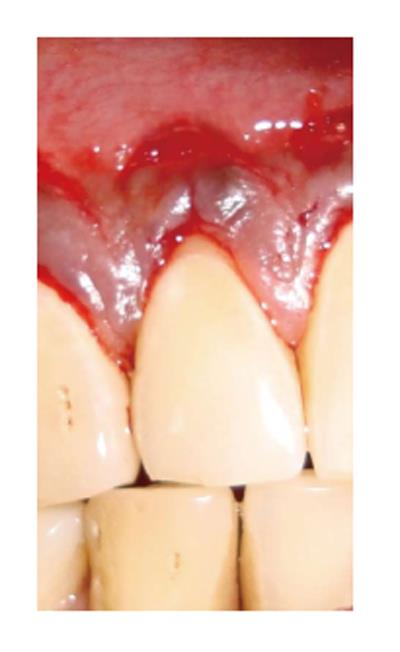
Figure 2 Coronally repositioned flap with 12.
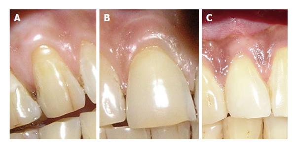
Figure 3 Six months post-operative view.
A: Six months post-operative view of 12; B: Six months post-operative view of 11; C: Six months post-operative view of 13.
- Citation: Mishra AK, Kumathalli K, Sridhar R, Maru R, Mangal B, Kedia S, Shrihatti R. Comparison of semilunar coronally repositioned flap with gingival massaging using an Ayurvedic product (irimedadi taila) in the treatment of class-I gingival recession: A clinical study. World J Clin Cases 2014; 2(10): 534-540
- URL: https://www.wjgnet.com/2307-8960/full/v2/i10/534.htm
- DOI: https://dx.doi.org/10.12998/wjcc.v2.i10.534


