Copyright
©The Author(s) 2024.
World J Clin Cases. May 16, 2024; 12(14): 2412-2419
Published online May 16, 2024. doi: 10.12998/wjcc.v12.i14.2412
Published online May 16, 2024. doi: 10.12998/wjcc.v12.i14.2412
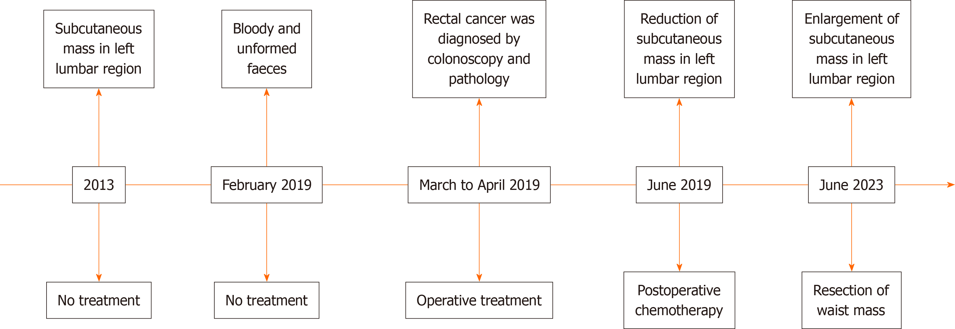
Figure 1 Timeline of the diagnosis and treatment of the patient.
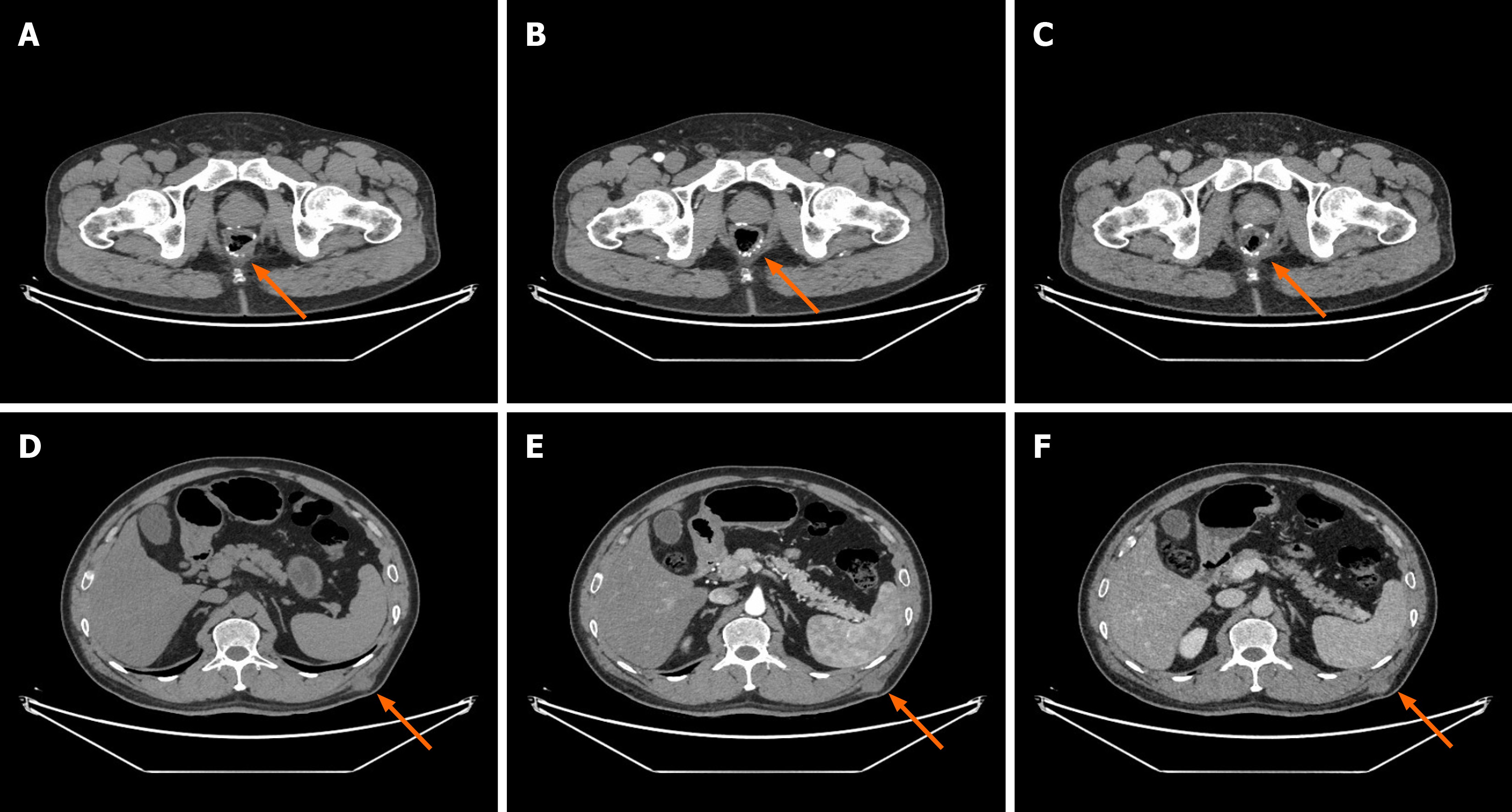
Figure 2 Contrast-enhanced abdominal computed tomography.
A: Rectal anastomosis plain scan; B: Rectal anastomosis arterial phase scan; C: Rectal anastomosis venous phase scan; D: Plain scan of the soft tissue mass of the left waist; E: Arterial phase scan of the soft tissue mass of the left waist; F: Venous phase scan of the soft tissue mass of the left waist.
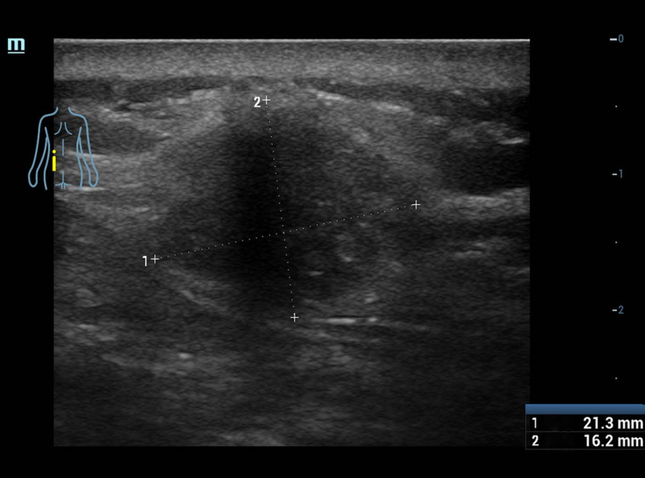
Figure 3 Ultrasound: A solid mass of 21 mm × 16 mm in size, irregularly bound and without obvious blood flow, located in the subcuta
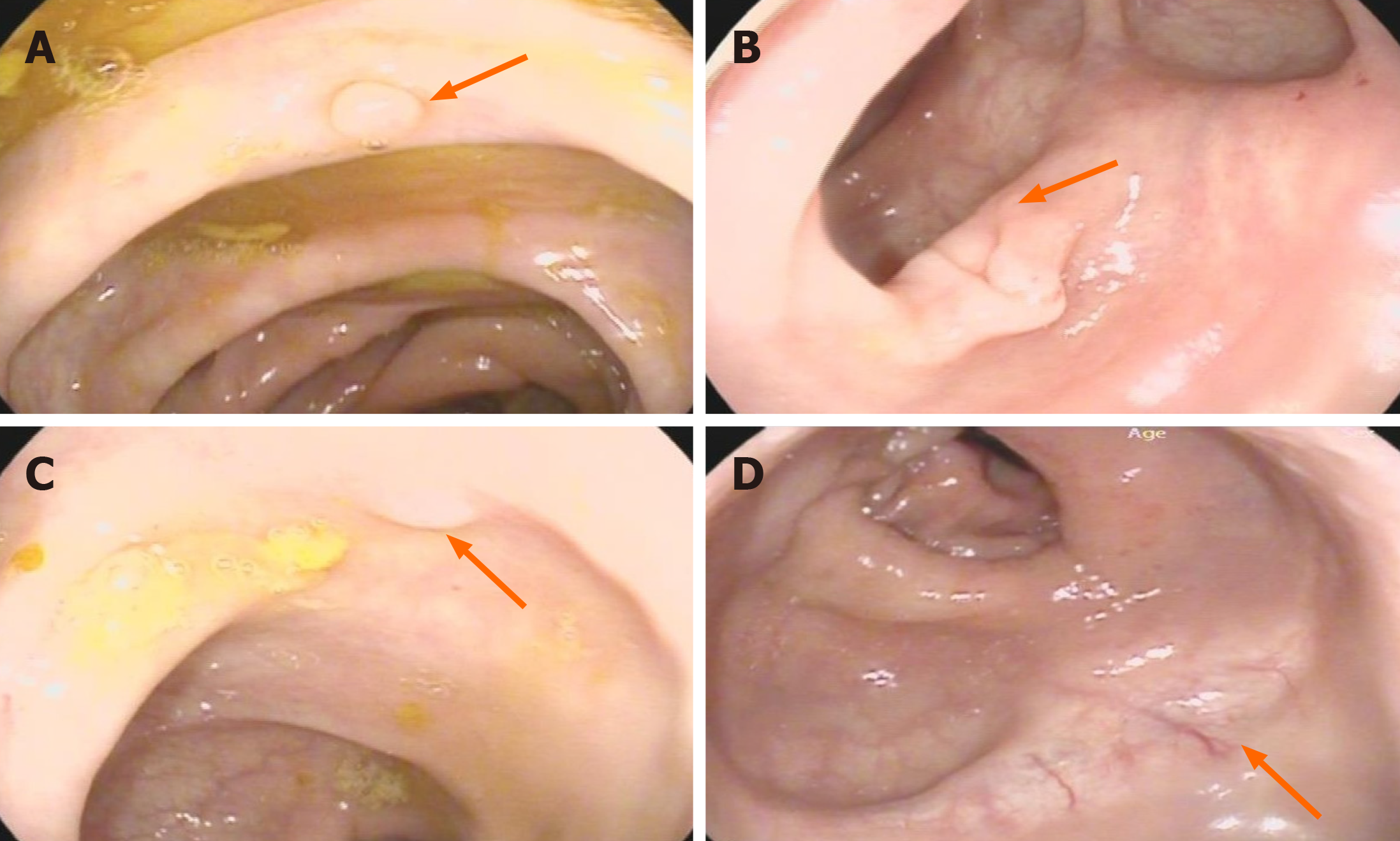
Figure 4 The colonoscopy results of this admission.
A: Low-grade tubular adenoma (ascending colon); B: Hyperplastic polyp (transverse colon); C: Hyperplastic polyp (sigmoid colon); D: Anastomotic scar.
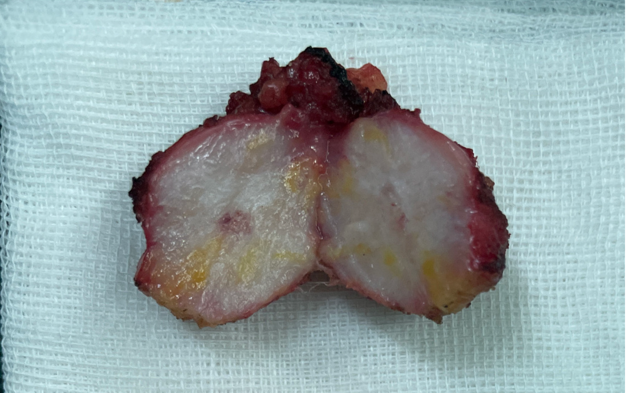
Figure 5 Soft tissue masses resected from the left waist subcutaneous soft tissue mass during surgery.
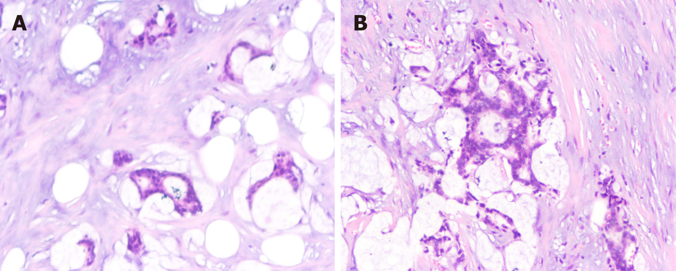
Figure 6 Pathological examination of the left waist mass.
A: Mucus is visible; B: Mucus is seen around adenocarcinoma cells (hematoxylin and eosin, × 200).
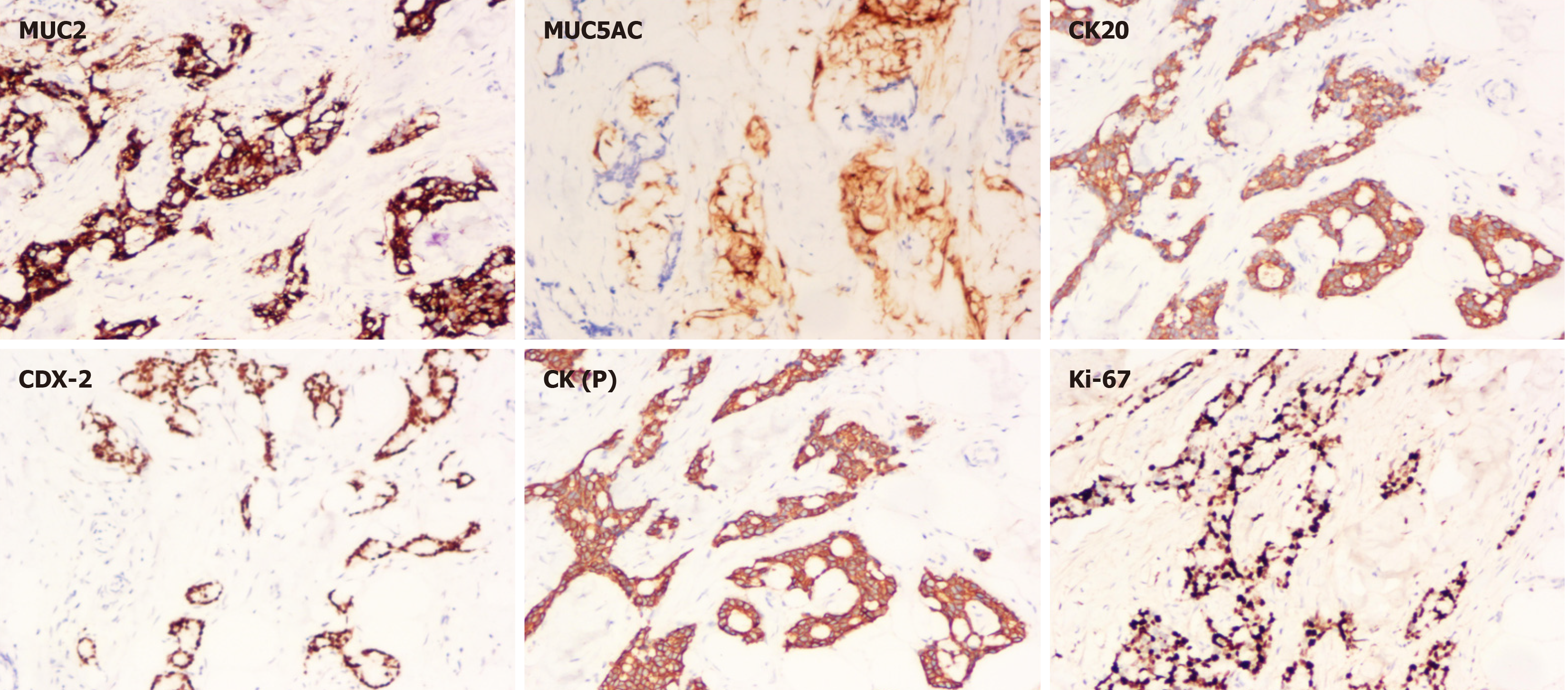
Figure 7 MUC2 (+), MUC5AC (partially +), CK20 (+), CDX-2 (+), CK (P) (+), Ki-67 (index 80%) (immunohistochemistry, × 200).
- Citation: Gong ZX, Li GL, Dong WM, Xu Z, Li R, Lv WX, Yang J, Li ZX, Xing W. Waist subcutaneous soft tissue metastasis of rectal mucinous adenocarcinoma: A case report. World J Clin Cases 2024; 12(14): 2412-2419
- URL: https://www.wjgnet.com/2307-8960/full/v12/i14/2412.htm
- DOI: https://dx.doi.org/10.12998/wjcc.v12.i14.2412


