Copyright
©The Author(s) 2024.
World J Clin Cases. Apr 16, 2024; 12(11): 1947-1953
Published online Apr 16, 2024. doi: 10.12998/wjcc.v12.i11.1947
Published online Apr 16, 2024. doi: 10.12998/wjcc.v12.i11.1947
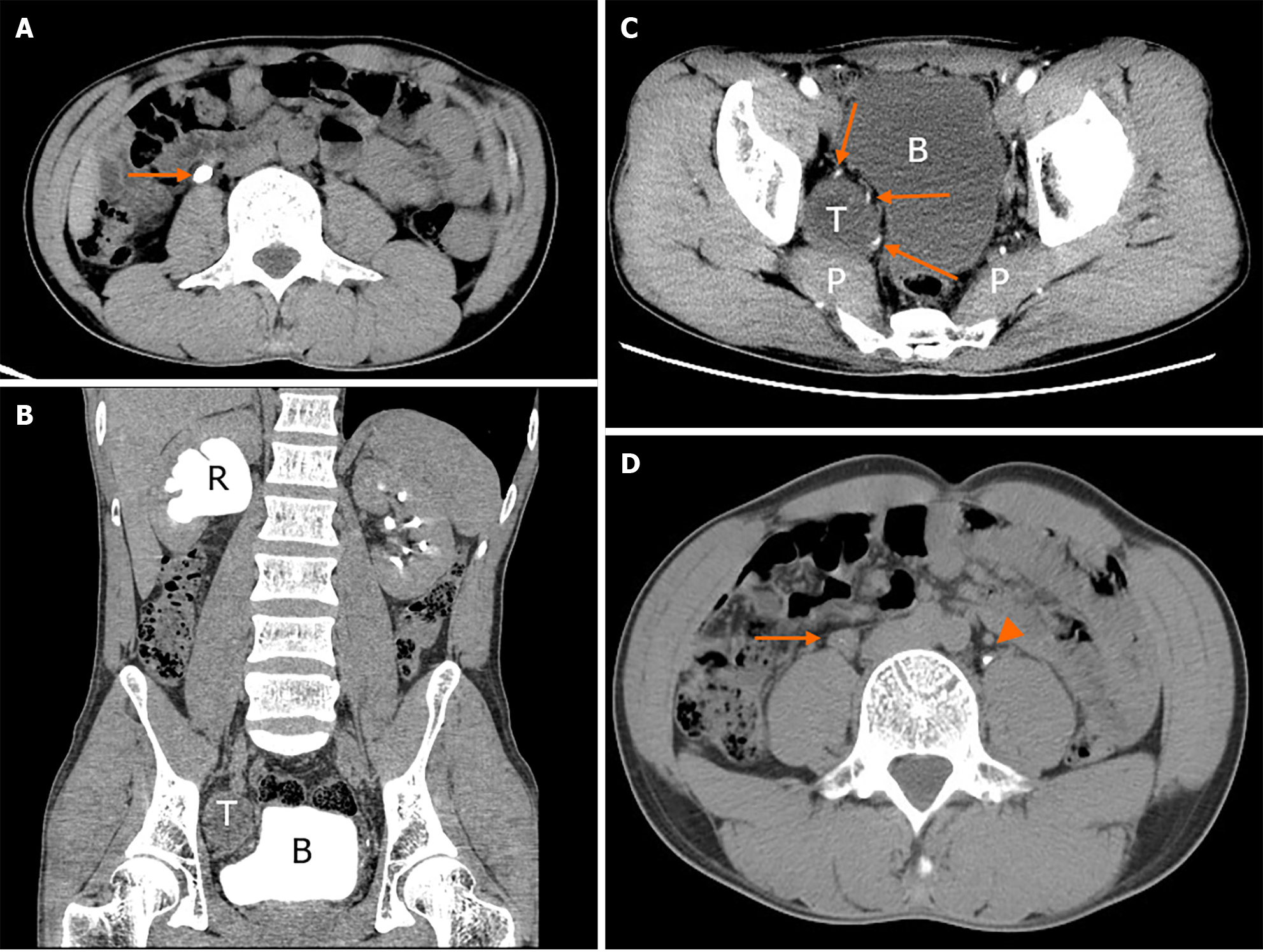
Figure 1 Abdominal and pelvic computed tomography scan.
A: Abdominal non-contrast axial computed tomography (CT) scan showed a 2-cm right upper ureteral calculi at the lumbar 3 level (arrow); B: The excretion phase of coronal CT urography (CTU) showed the dilated hydronephrotic renal pelvis caused by right ureteral stone obstruction and the deformed bladder caused by pelvic tumor compression; C: Contrast axial CT scan showed the anatomical location of pelvic tumor with blurred boundary between the posterior part of the mass and the piriformis muscle and abundant arterial supply at the anterior, medial and posterior part of the mass (arrows); D: The excretion phase of axial CTU showed the ureter (arrow) beneath the stone was still obviously dilated compared with the contralateral ureter (arrowhead). R: Renal pelvis; T: Pelvic tumor; P: Piriformis muscle; B: Bladder.
Figure 2 Immunohistochemical staining and evaluation.
A: The histopathological section from pelvic tumor showed typical characteristics of alternating Antoni B and Antoni A with spindle tumor cells arranged in palisade or whorl pattern (hematoxylin and eosin, × 100); B and C: Immunohistochemical detection showed diffuse strong S100 expression and sporadic Ki-67 expression (arrows) (× 100). a: Antoni A; b: Antoni B.
- Citation: Xiong Y, Li J, Yang HJ. Concomitant treatment of ureteral calculi and ipsilateral pelvic sciatic nerve schwannoma with transperitoneal laparoscopic approach: A case report. World J Clin Cases 2024; 12(11): 1947-1953
- URL: https://www.wjgnet.com/2307-8960/full/v12/i11/1947.htm
- DOI: https://dx.doi.org/10.12998/wjcc.v12.i11.1947


