Copyright
©The Author(s) 2024.
World J Clin Cases. Apr 16, 2024; 12(11): 1909-1917
Published online Apr 16, 2024. doi: 10.12998/wjcc.v12.i11.1909
Published online Apr 16, 2024. doi: 10.12998/wjcc.v12.i11.1909
Figure 1 Possible etiopathogenesis of splenic hamartoma.
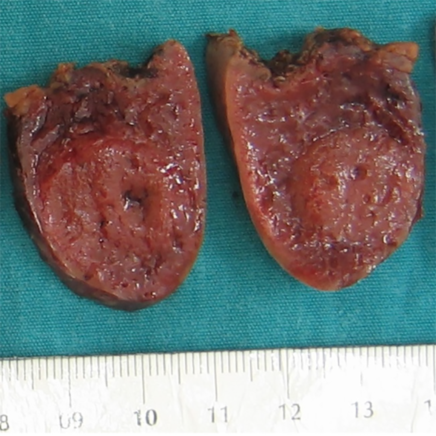
Figure 2 Gross appearance of splenic hamartoma.
A nodular circumscribed unencapsulated lesion is visible on the cross-section, representing splenic hamartoma.
Figure 3 Microscopic images of the splenic hamartoma.
A: Splenic hamartoma adjacent to normal splenic parenchyma was observed in the upper left of the panel, and compressed splenic parenchyma was observed in the lower right [hematoxylin and eosin (HE) × 50]; B: The lesion was composed of disorganized vascular channels lined by endothelial cells without significant cytological atypia (HE × 200); C: No mitosis or atypical cells were observed (HE × 400).
Figure 4 Immunohistochemical staining of the splenic hamartoma.
A: CD8 was positive in the lining cells of vascular channels and in rare lymphocytes (× 200); B: Strong staining for CD31 was observed in the lesion (× 200); C: The lining cells of sinus-like spaces in the hamartoma were immunoreactive for CD34 (× 200).
- Citation: Milickovic M, Rasic P, Cvejic S, Bozic D, Savic D, Mijovic T, Cvetinovic S, Djuricic SM. Splenic hamartomas in children. World J Clin Cases 2024; 12(11): 1909-1917
- URL: https://www.wjgnet.com/2307-8960/full/v12/i11/1909.htm
- DOI: https://dx.doi.org/10.12998/wjcc.v12.i11.1909


