Copyright
©The Author(s) 2023.
World J Clin Cases. Mar 16, 2023; 11(8): 1830-1836
Published online Mar 16, 2023. doi: 10.12998/wjcc.v11.i8.1830
Published online Mar 16, 2023. doi: 10.12998/wjcc.v11.i8.1830
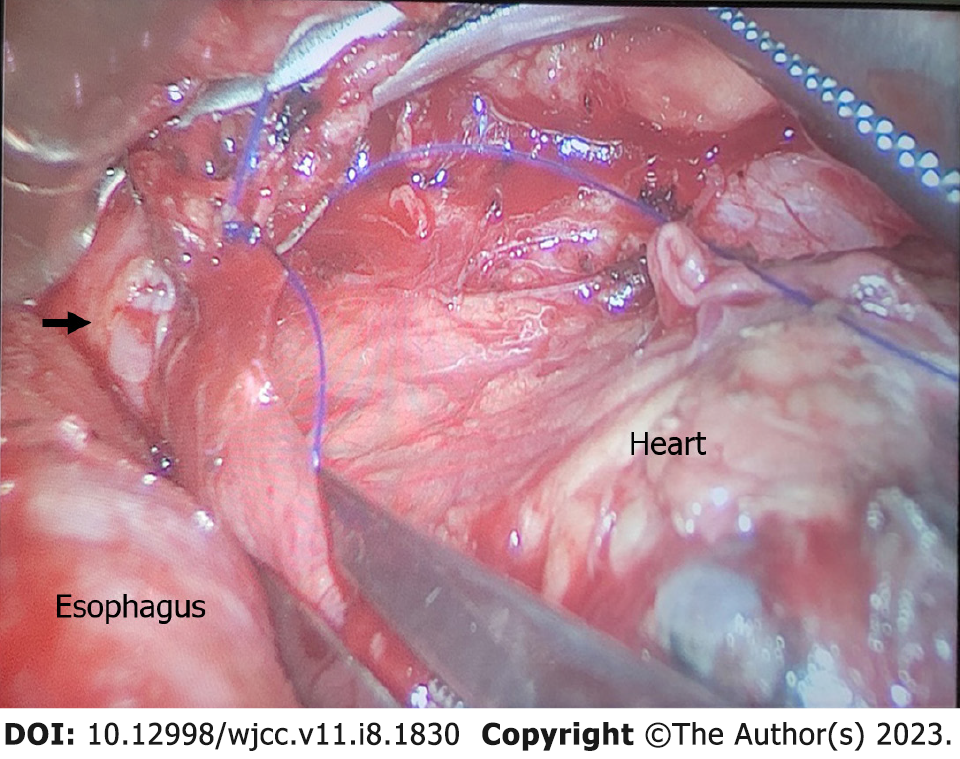
Figure 1 After occlusion of the left inferior pulmonary vein.
The arrow indicates the vascular rupture.
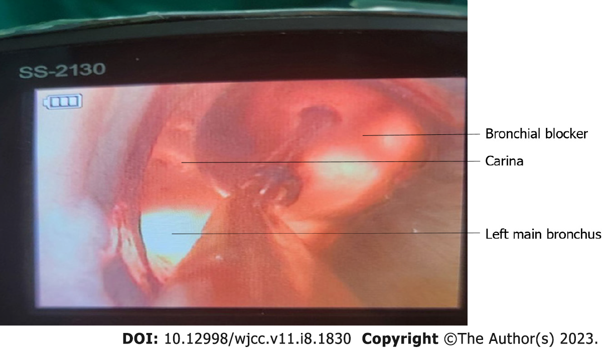
Figure 2
Bronchial blocker in the correct position.
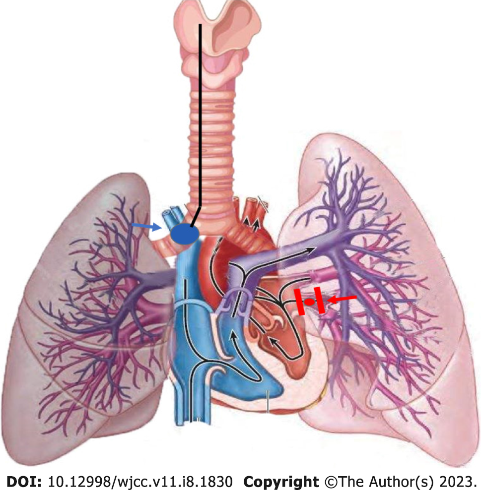
Figure 3
The blue arrow indicates the bronchial blocker and the red arrow the bleeding point and forceps.
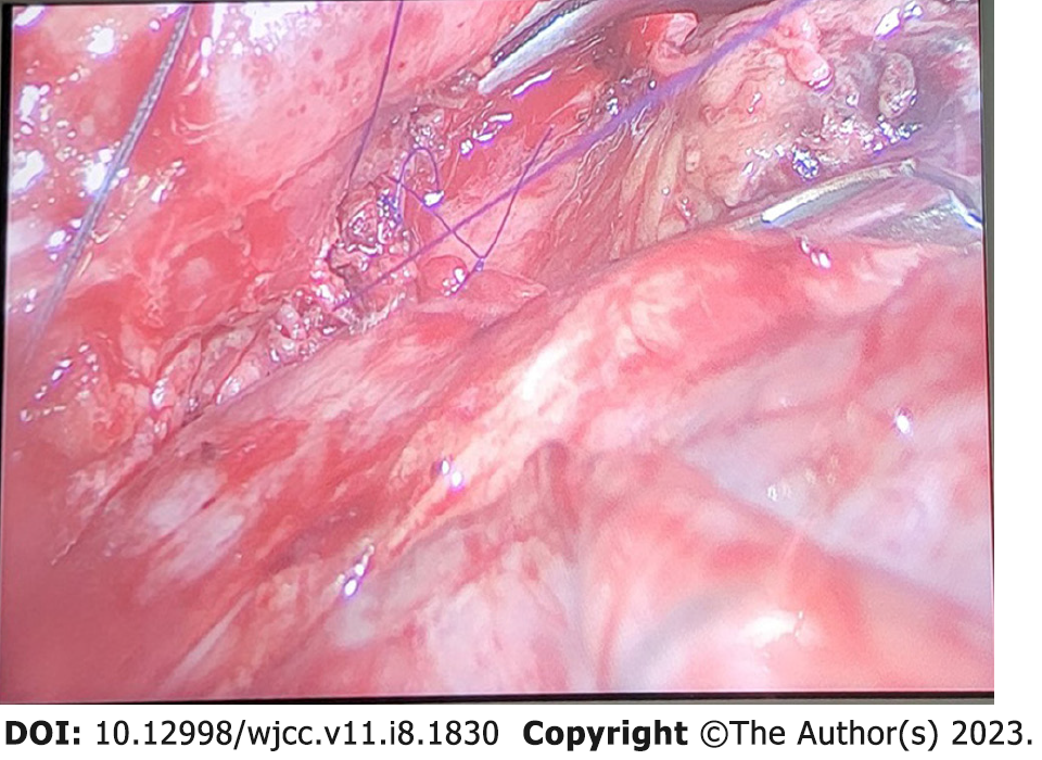
Figure 4
Blood vessels after suture.
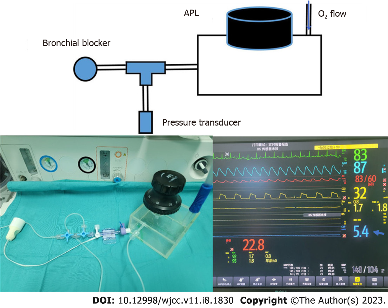
Figure 5 Accurate control of the continuous positive airway pressure and physical diagrams.
The blue arrow indicates the continuous positive airway pressure displayed in the monitor.
- Citation: Zhou C, Song S, Fu JF, Zhao XL, Liu HQ, Pei HS, Guo HB. Continuous positive airway pressure for treating hypoxemia due to pulmonary vein injury: A case report. World J Clin Cases 2023; 11(8): 1830-1836
- URL: https://www.wjgnet.com/2307-8960/full/v11/i8/1830.htm
- DOI: https://dx.doi.org/10.12998/wjcc.v11.i8.1830


