Copyright
©The Author(s) 2023.
World J Clin Cases. Dec 16, 2023; 11(35): 8411-8415
Published online Dec 16, 2023. doi: 10.12998/wjcc.v11.i35.8411
Published online Dec 16, 2023. doi: 10.12998/wjcc.v11.i35.8411
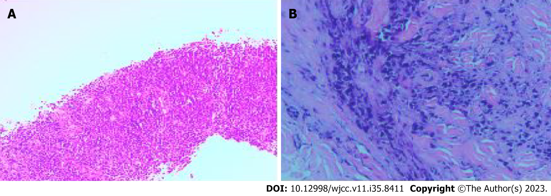
Figure 1 Pathological findings.
Pathological examination revealed poorly differentiated adenocarcinoma. A: Biopsy of cervical lymph node; B: Skin biopsy of the chest wall.
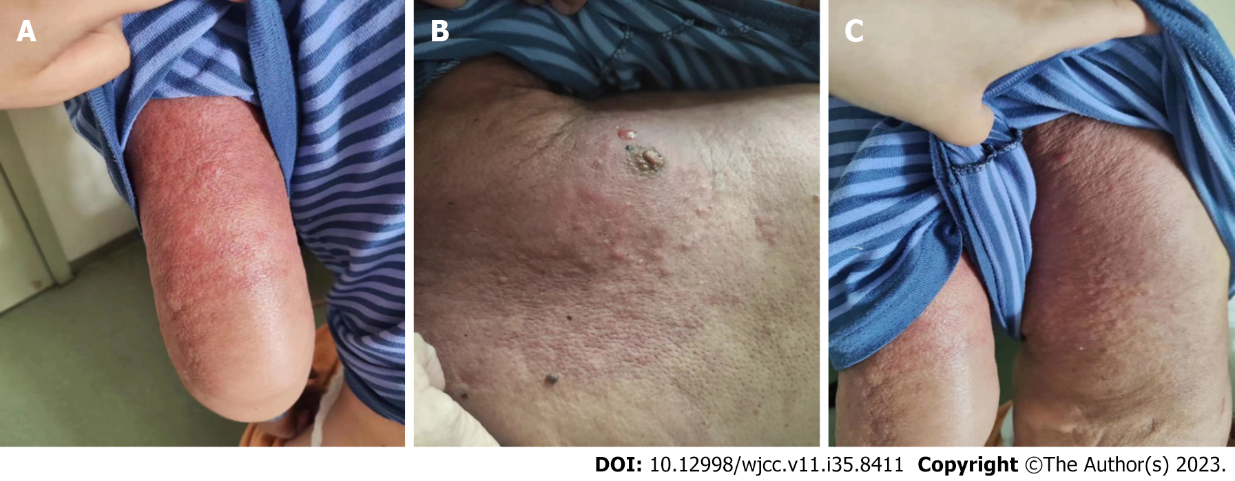
Figure 2 Cutaneous metastases from gastric cancer.
The extensive skin redness and swelling, accompanied by skin thickening and scattered small nodules. This image is published with the patient’s guardian consent. A: Left upper limb; B: Left chest wall; C: Left back.
- Citation: Tian L, Ye ZB, Du YL, Li QF, He LY, Zhang HZ. Inflammatory cutaneous metastases originating from gastric cancer: A case report. World J Clin Cases 2023; 11(35): 8411-8415
- URL: https://www.wjgnet.com/2307-8960/full/v11/i35/8411.htm
- DOI: https://dx.doi.org/10.12998/wjcc.v11.i35.8411


