Copyright
©The Author(s) 2023.
World J Clin Cases. Dec 16, 2023; 11(35): 8343-8349
Published online Dec 16, 2023. doi: 10.12998/wjcc.v11.i35.8343
Published online Dec 16, 2023. doi: 10.12998/wjcc.v11.i35.8343
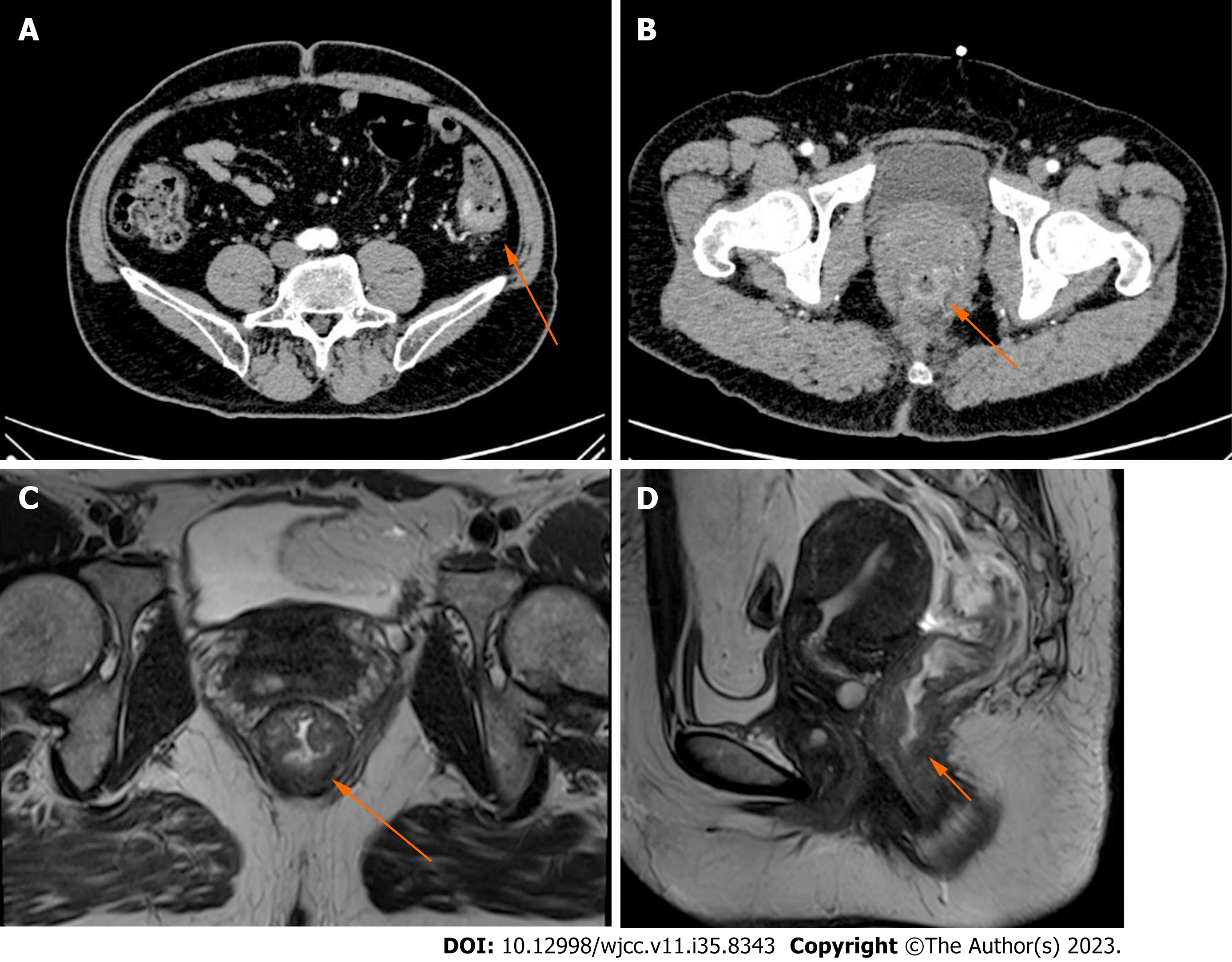
Figure 1 Computed tomography images.
A: Abdominal computed tomography (CT) revealed intestinal stenosis in the splenic flexure of the colon (arrow); B: Abdominal CT revealed obvious bowel wall thickness in the lower portions of the rectum (arrow); C and D: Axial and sagittal magnetic resonance imaging of the rectum depicted a sign of concentric tumor rings within the rectal wall and a tumor penetrating the layer of the intrinsic muscle into the perirectal mesenteric fat (arrow).
Figure 2 Colonoscopic images.
A: An irregularly shaped neoplasm in the right rectal wall occupying over 1/3 of the rectum circumference approximately 2 cm from the anal margin; B: A tumor-like lesion causing intestinal stenosis in the descending colon occupying 1/2 of the colon circumference approximately 32 cm from the anal margin; C: A 2 cm wide polypoidal neoplasm in the ileocecum.
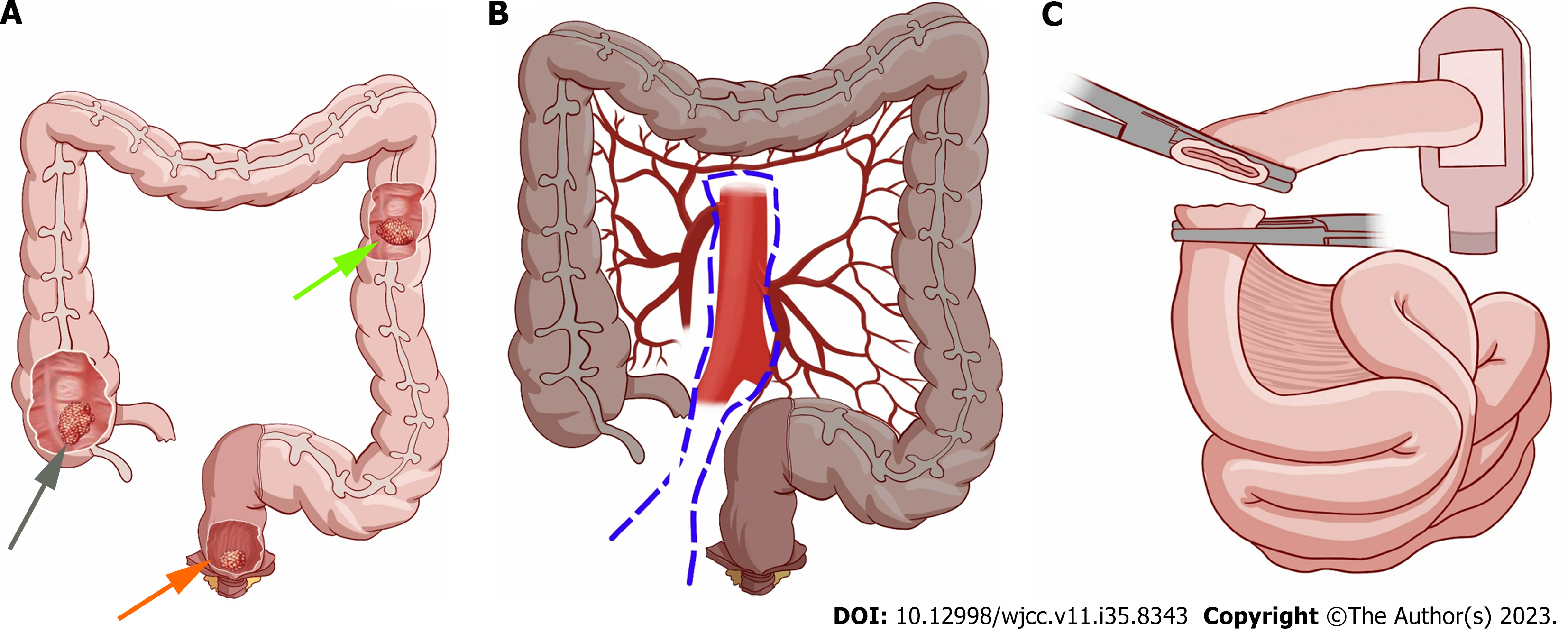
Figure 3 Diagram of location of three different tumors and extent of resection for this patient.
A: Three different tumors are located in the ileocecum (gray arrow), descending colon (green arrow), and lower portions of rectum (orange arrow), respectively; B: Extent of resection: Left hemicolon, right hemicolon, sigmoid colon, rectum, and anal canal were removed (dark section), including mesentery, blood vessels, and lymph nodes; C: Ileostomy was performed.
Figure 4 Pathological diagnosis (magnification 10 × 10).
A: Mucinous adenocarcinoma in the rectum. The mass invaded into the muscular layer; B: Moderately differentiated adenocarcinoma in the descending colon. The mass invaded into the muscular layer; C: Well-differentiated adenocarcinoma in the ascending colon. The mass invaded the submucosal layer.
- Citation: Li F, Zhao B, Zhang L, Chen GQ, Zhu L, Feng XL, Yao H, Tang XF, Yang H, Liu YQ. Rare synchronous colorectal carcinoma with three pathological subtypes: A case report and review of the literature. World J Clin Cases 2023; 11(35): 8343-8349
- URL: https://www.wjgnet.com/2307-8960/full/v11/i35/8343.htm
- DOI: https://dx.doi.org/10.12998/wjcc.v11.i35.8343


