Copyright
©The Author(s) 2023.
World J Clin Cases. Jan 16, 2023; 11(2): 464-471
Published online Jan 16, 2023. doi: 10.12998/wjcc.v11.i2.464
Published online Jan 16, 2023. doi: 10.12998/wjcc.v11.i2.464
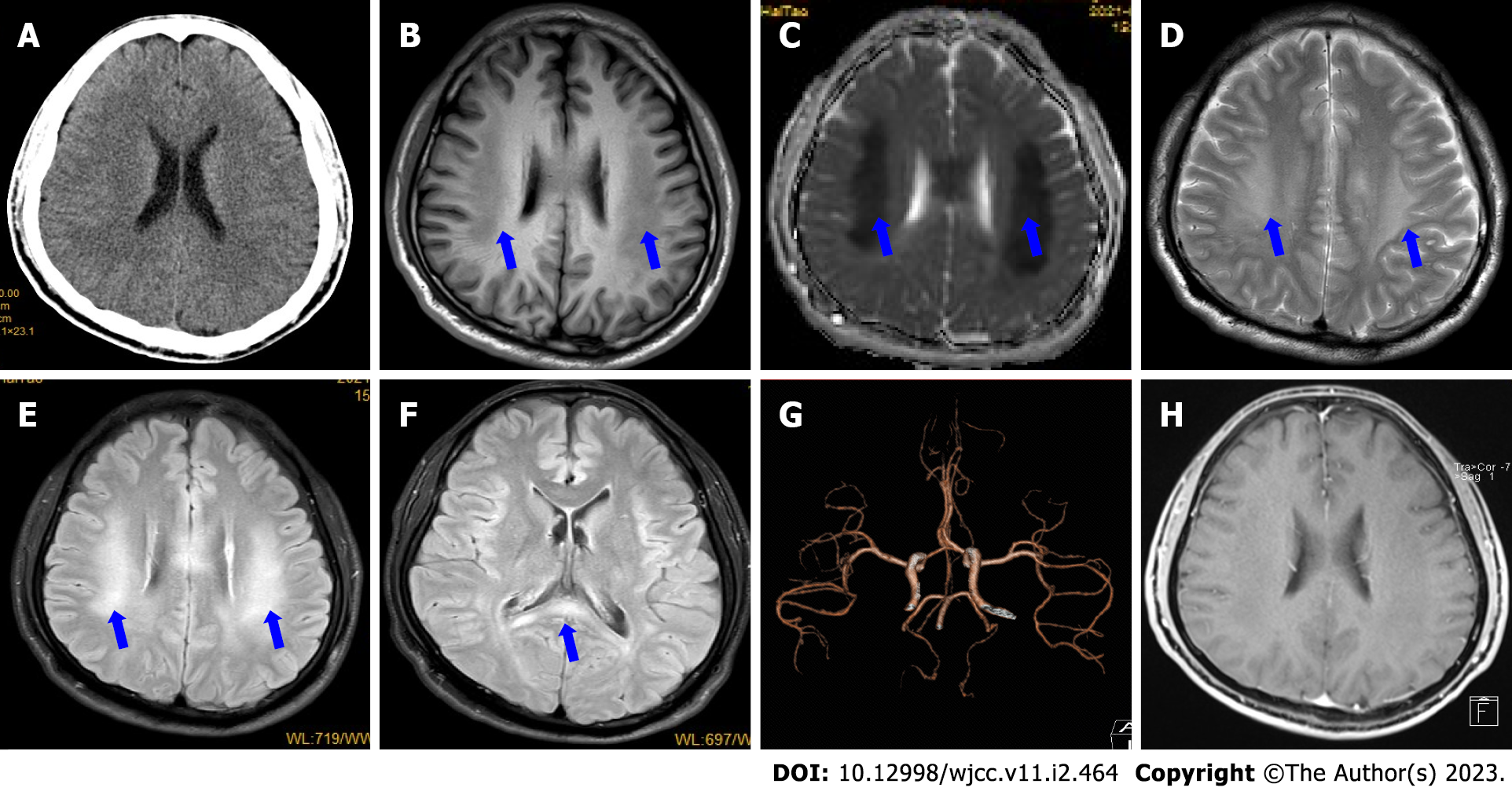
Figure 1 Initial magnetic resonance imaging findings.
A: Initial head computed tomography (CT) shows no obvious abnormalities; B: Magnetic resonance imaging (MRI) on day 2 showed bilateral, symmetric, and definite hypointensities with T1-weighted imaging (blue arrows); C and D: Hypointensities on apparent diffusion coefficient mapping (C) and T2-weighted imaging (D) (blue arrows); E and F: Hyperintensities on fluid-attenuation inversion recovery sequence in the corpus callosum and supratentorial white matter (blue arrows); G: CT angiography shows no obvious abnormalities; H: Contrast-enhanced MRI on day 4 shows that the lesions were not enhanced.
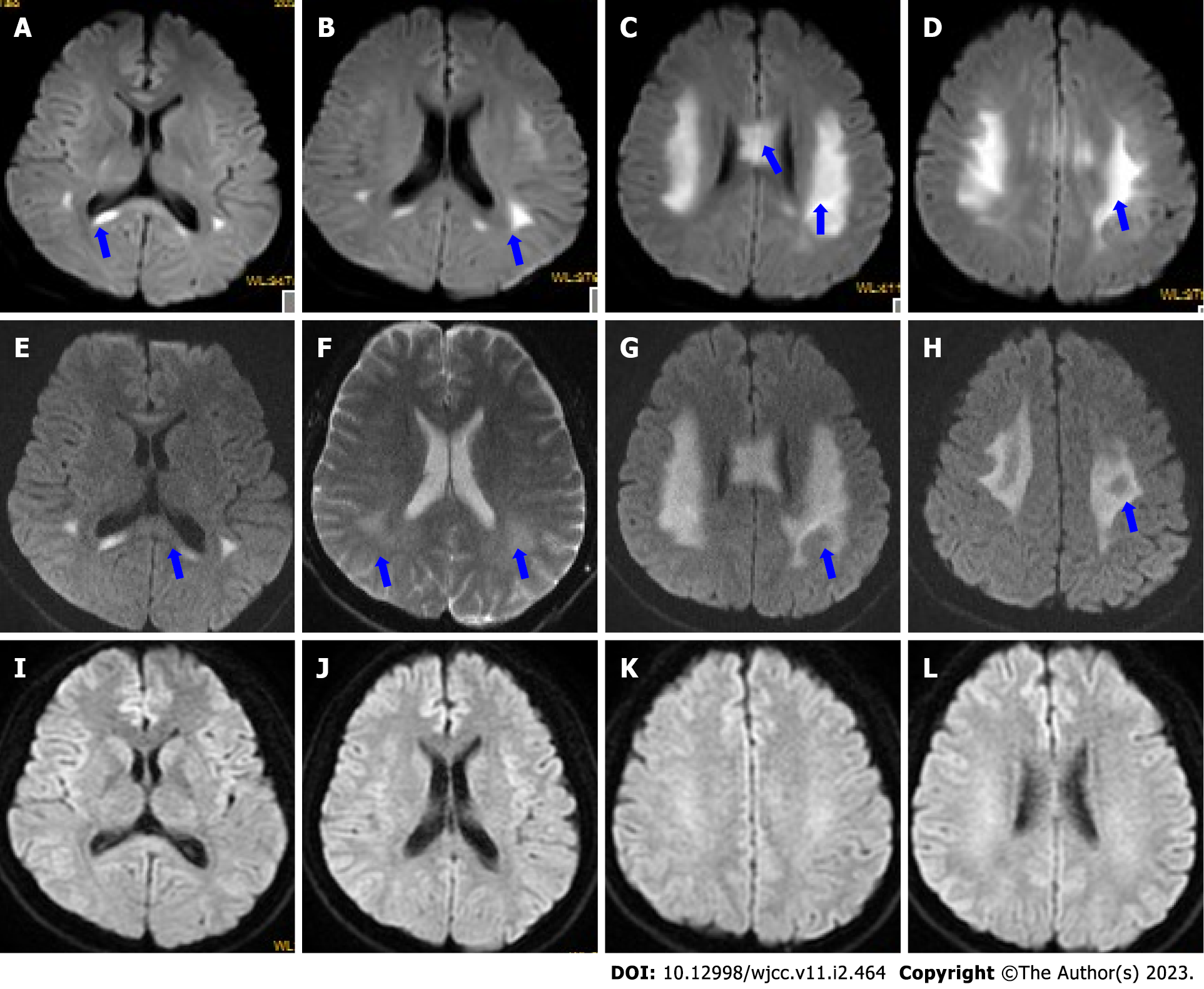
Figure 2 Dynamic changes in diffusion-weighted imaging.
A-D: Diffusion-weighted imaging (DWI) on day 2 showed that the corpus callosum (A-C) and bilateral centrum semiovale (B-D) had marked abnormally restricted diffusion (blue arrows); E-H: DWI on day 4 showed that the areas of restricted diffusion were obvious (F and G) but not reduced (blue arrows); I-L: DWI at 1.5 mo after discharge shows that the lesions disappeared.
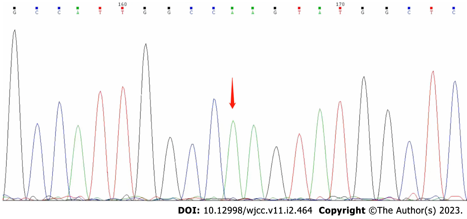
Figure 3 Genetic sequencing (Beijing Golden Standard Medical Laboratory, Beijing, China) results showed a hemizygous mutation (c.
65 G>A p.R22Q) in the GJB1 gene.
- Citation: Zhang Q, Wang Y, Bai RT, Lian BR, Zhang Y, Cao LM. X-linked Charcot-Marie-Tooth disease after SARS-CoV-2 vaccination mimicked stroke-like episodes: A case report. World J Clin Cases 2023; 11(2): 464-471
- URL: https://www.wjgnet.com/2307-8960/full/v11/i2/464.htm
- DOI: https://dx.doi.org/10.12998/wjcc.v11.i2.464


