Copyright
©The Author(s) 2023.
World J Clin Cases. Jan 6, 2023; 11(1): 249-254
Published online Jan 6, 2023. doi: 10.12998/wjcc.v11.i1.249
Published online Jan 6, 2023. doi: 10.12998/wjcc.v11.i1.249
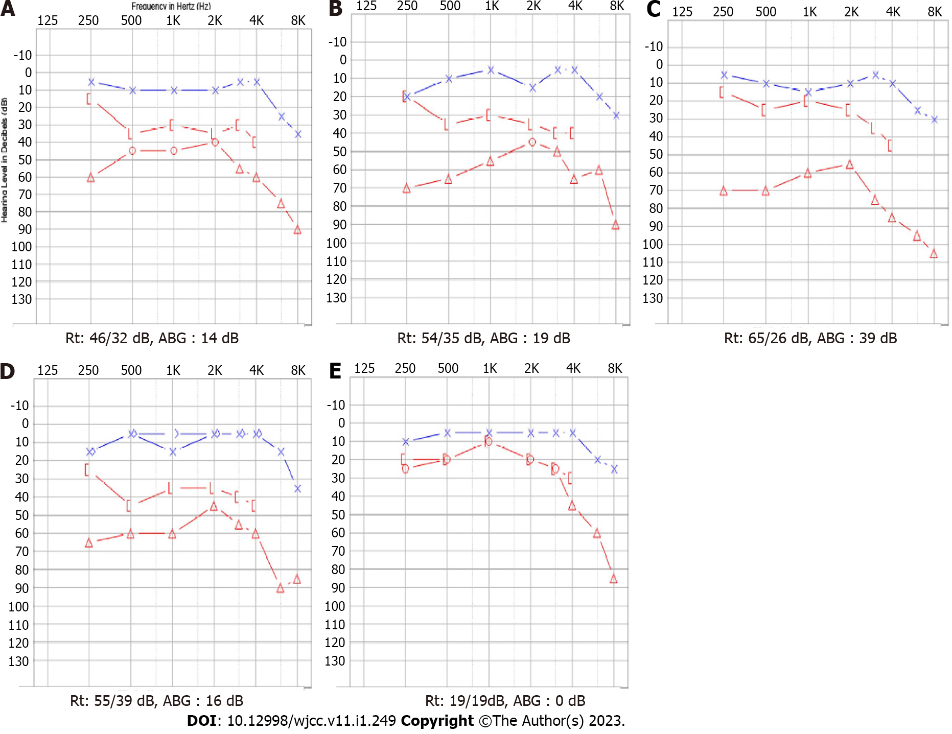
Figure 1 Pure tone audiogram.
A: At the initial visit, a pure-tone audiogram showed moderate mixed hearing loss on the right ear [bond conduction (BC) 32 dB hearing level (HL), air conduction (AC) 46 dB HL] with an air-bone gap (ABG) of 14 dB, whereas the left ear showed a normal HL; B: At two months after the initial visit, the pure tone threshold on the right was (BC 35 dB HL, AC 54 dB HL) with an ABG of 19 dB; C: At revisit after 2.5 years, a pure-tone audiogram showed moderate to severe mixed hearing loss on the right (BC 26 dB HL, AC 65 dB HL) with an ABG of 39 dB; D: A pure tone auditogram one day before right exploratory tympanotomy showed moderate mixed hearing loss on the right (BC 39 dB HL, AC 55 dB HL) with an ABG of 16 dB; E: A pure-tone audiogram conducted 1 mo after the surgery showed a normal HL (BC 19 dB HL, AC 19 dB HL) with no ABG on the right ear. ABG: Air-bone gap.
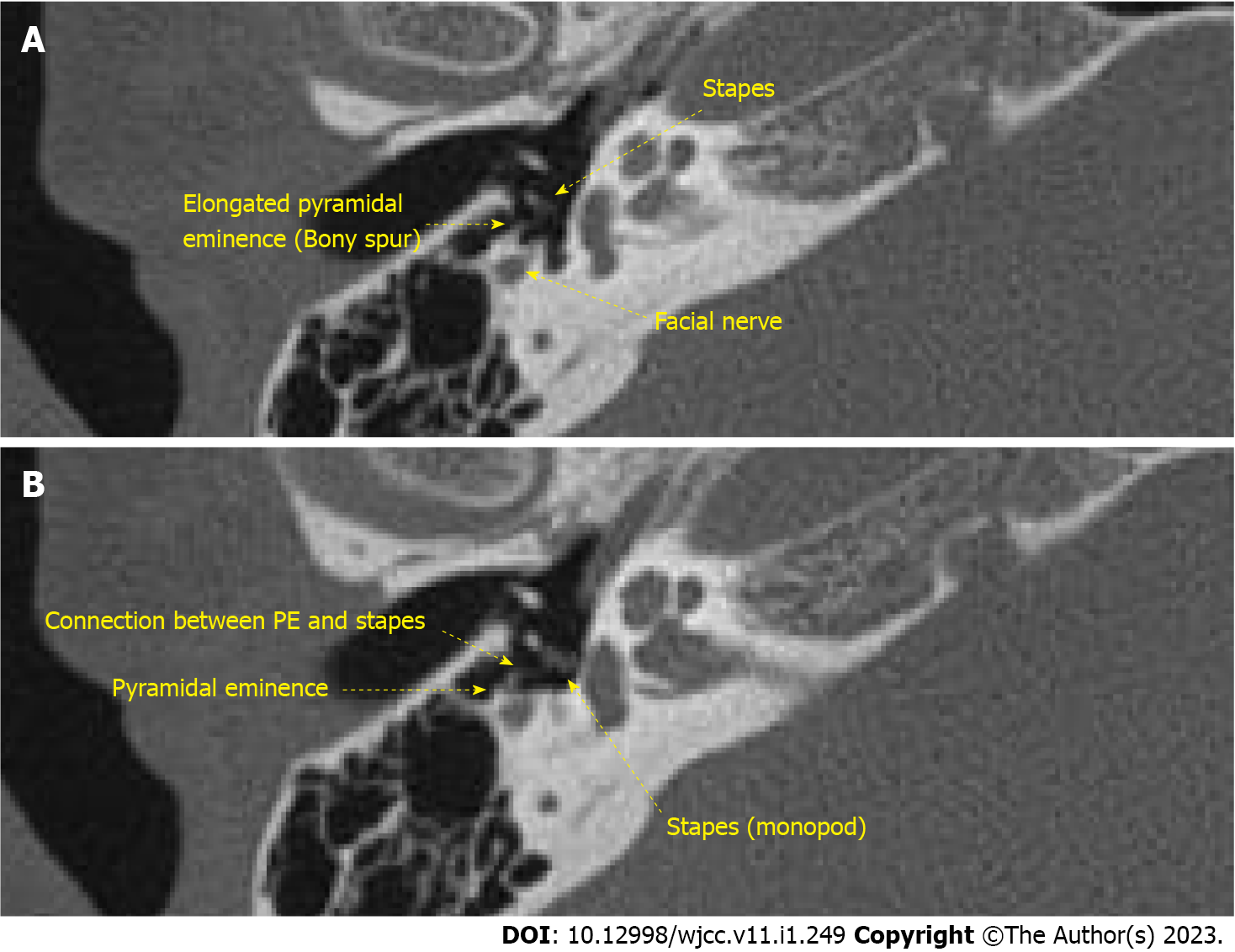
Figure 2 Temporal bone computed tomography.
A: Elongated pyramidal eminence (PE), facial nerve, and stapes are shown (yellow arrows); B: PE, connection between PE and stapes and monopod stapes are noted (yellow arrows). PE: Pyramidal eminence.
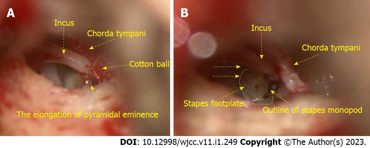
Figure 3 Microscopic examination.
A: In the operation, elongation of pyramidal eminence was noted, and it was connected to the stapes head; B: The stapes crura was thickened by the fusion of anterior and posterior crura creating a monopod shape.
- Citation: Choi S, Park SH, Kim JS, Chang J. Congenital stapes suprastructure fixation presenting with fluctuating auditory symptoms: A case report. World J Clin Cases 2023; 11(1): 249-254
- URL: https://www.wjgnet.com/2307-8960/full/v11/i1/249.htm
- DOI: https://dx.doi.org/10.12998/wjcc.v11.i1.249


