Copyright
©The Author(s) 2023.
World J Clin Cases. Jan 6, 2023; 11(1): 150-156
Published online Jan 6, 2023. doi: 10.12998/wjcc.v11.i1.150
Published online Jan 6, 2023. doi: 10.12998/wjcc.v11.i1.150
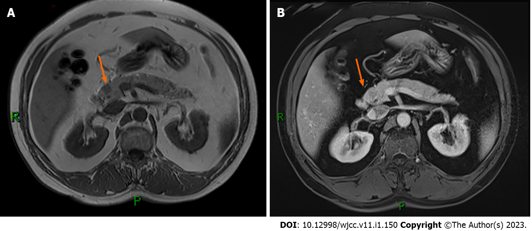
Figure 1 Small and slightly long T1 signal nodules were seen in the head of the pancreas with clear boundaries (A) and mild enhancement (B).
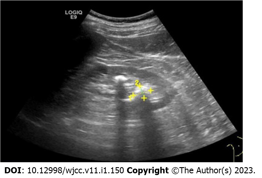
Figure 2 Endoscopic ultrasonography suggested a hypoechoic lesion in the head of the pancreas, with a hyperechoic nodule, about 1 cm × 1.
4 cm in size.
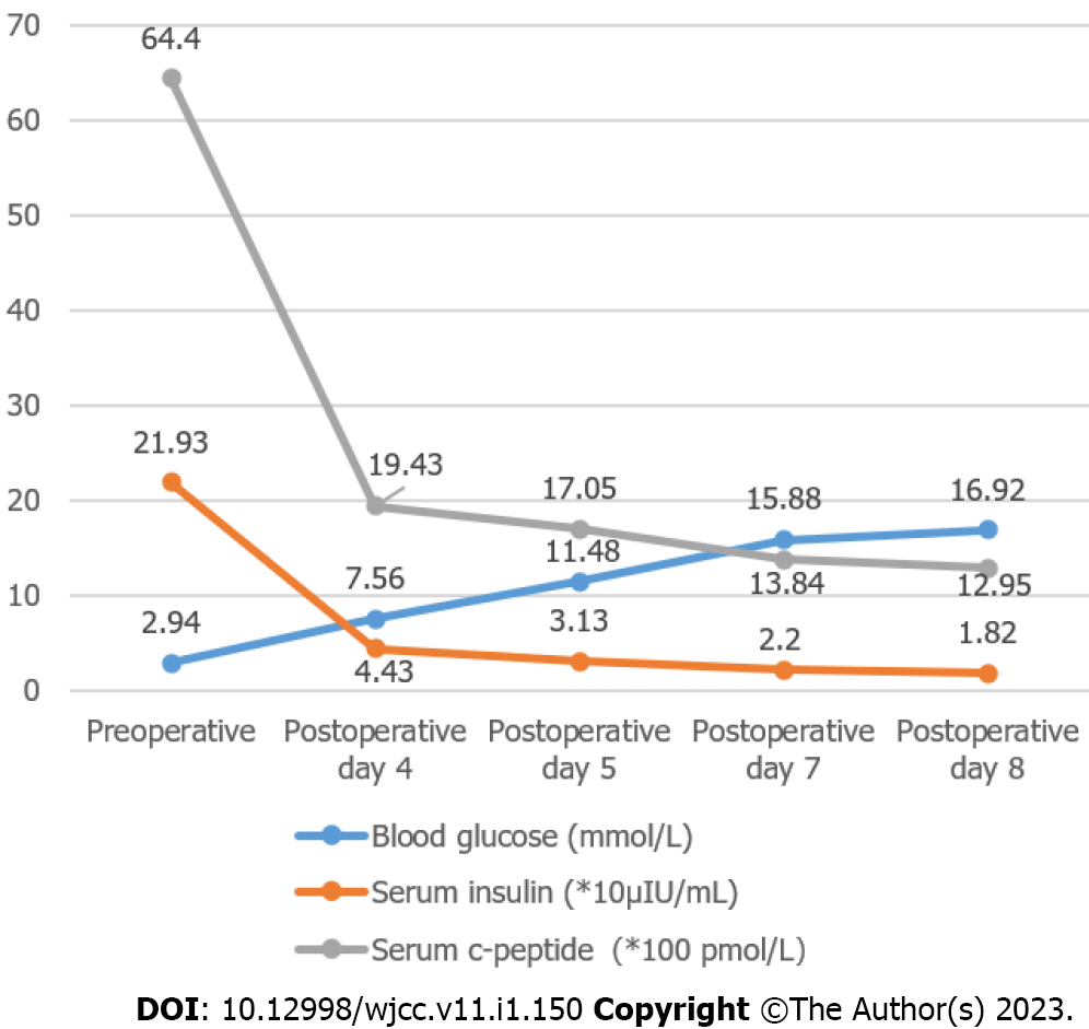
Figure 3 Preoperative and postoperative changes of fasting blood glucose, insulin and C-peptide.
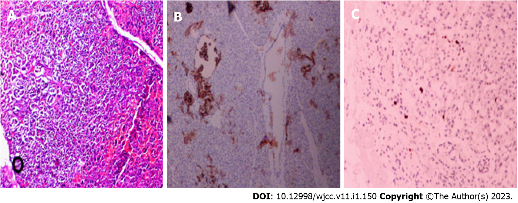
Figure 4 Postoperative pathological results showed nesidioblastosis.
A: Islet cells show hypertrophy, with pleomorphic changes in the nucleus, an increased and transparent cytoplasm; and immunohistochemistry analysis showed Ki-67 about 2% (+) (B), Syn (+)(C), MGMT (+), CD56 (-), CgA (-), Insulin (-), CK(+).
- Citation: Tu K, Zhao LJ, Gu J. Adult focal β-cell nesidioblastosis: A case report. World J Clin Cases 2023; 11(1): 150-156
- URL: https://www.wjgnet.com/2307-8960/full/v11/i1/150.htm
- DOI: https://dx.doi.org/10.12998/wjcc.v11.i1.150


