Copyright
©The Author(s) 2022.
World J Clin Cases. Oct 26, 2022; 10(30): 11198-11203
Published online Oct 26, 2022. doi: 10.12998/wjcc.v10.i30.11198
Published online Oct 26, 2022. doi: 10.12998/wjcc.v10.i30.11198
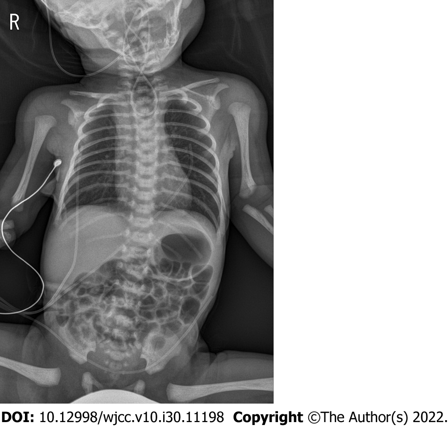
Figure 1 An infantogram of the neonate.
The tip of the catheter is shown to be curled up in the upper chest and neck area due to esophageal atresia.
Figure 2 Tracheal computed tomography of the neonate.
A: Sagittal image. The measured size of the fistula is 6.60 mm (red bidirectional arrow). The trachea and esophagus are at an obtuse angle of approximately 146° (orange lines). The measured length from the lip to the carina to predict the appropriate depth of the endotracheal tube is approximately 9.8 cm (yellow dashed line); B: Coronal image. The measured size of the fistula is 4.54 mm (red bidirectional arrow). The fistula is 10.2 mm above the carina (orange bidirectional arrow). T: Trachea; E: Esophagus; O: Opening of the fistula; C: Carina.
- Citation: Hwang SM, Kim MJ, Kim S, Kim S. Accidental esophageal intubation via a large type C congenital tracheoesophageal fistula: A case report. World J Clin Cases 2022; 10(30): 11198-11203
- URL: https://www.wjgnet.com/2307-8960/full/v10/i30/11198.htm
- DOI: https://dx.doi.org/10.12998/wjcc.v10.i30.11198


