Copyright
©The Author(s) 2022.
World J Clin Cases. Oct 26, 2022; 10(30): 11090-11100
Published online Oct 26, 2022. doi: 10.12998/wjcc.v10.i30.11090
Published online Oct 26, 2022. doi: 10.12998/wjcc.v10.i30.11090
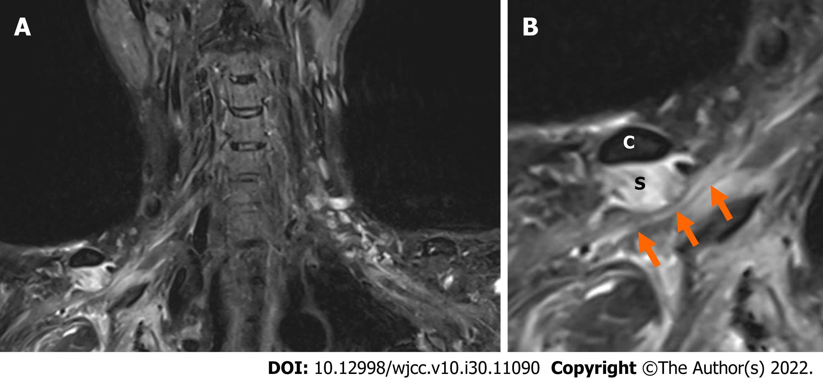
Figure 1 A T2-weighted magnetic resonance coronal image of the brachial plexus.
A: A reduced version; B: An enlarged version. The hypertrophied subclavius muscle is compressing the underlying brachial plexus, and elevated signal intensity throughout the brachial plexus is shown (arrows). C: Clavicle; S: Subclavius muscle.
Figure 2 Musculoskeletal ultrasonography along the short axis of the subclavius muscle.
A: Isolated hypertrophy of subclavius muscle (arrowhead) of the affected side; B: Normal finding of subclavius muscle (arrowhead) of the unaffected side.
Figure 3 A photograph of the patient showing the incision site of the surgery.
- Citation: Go YI, Kim DS, Kim GW, Won YH, Park SH, Ko MH, Seo JH. Recovery of brachial plexus injury after bronchopleural fistula closure surgery based on electrodiagnostic study: A case report and review of literature. World J Clin Cases 2022; 10(30): 11090-11100
- URL: https://www.wjgnet.com/2307-8960/full/v10/i30/11090.htm
- DOI: https://dx.doi.org/10.12998/wjcc.v10.i30.11090


