Copyright
©The Author(s) 2022.
World J Clin Cases. Oct 26, 2022; 10(30): 11082-11089
Published online Oct 26, 2022. doi: 10.12998/wjcc.v10.i30.11082
Published online Oct 26, 2022. doi: 10.12998/wjcc.v10.i30.11082
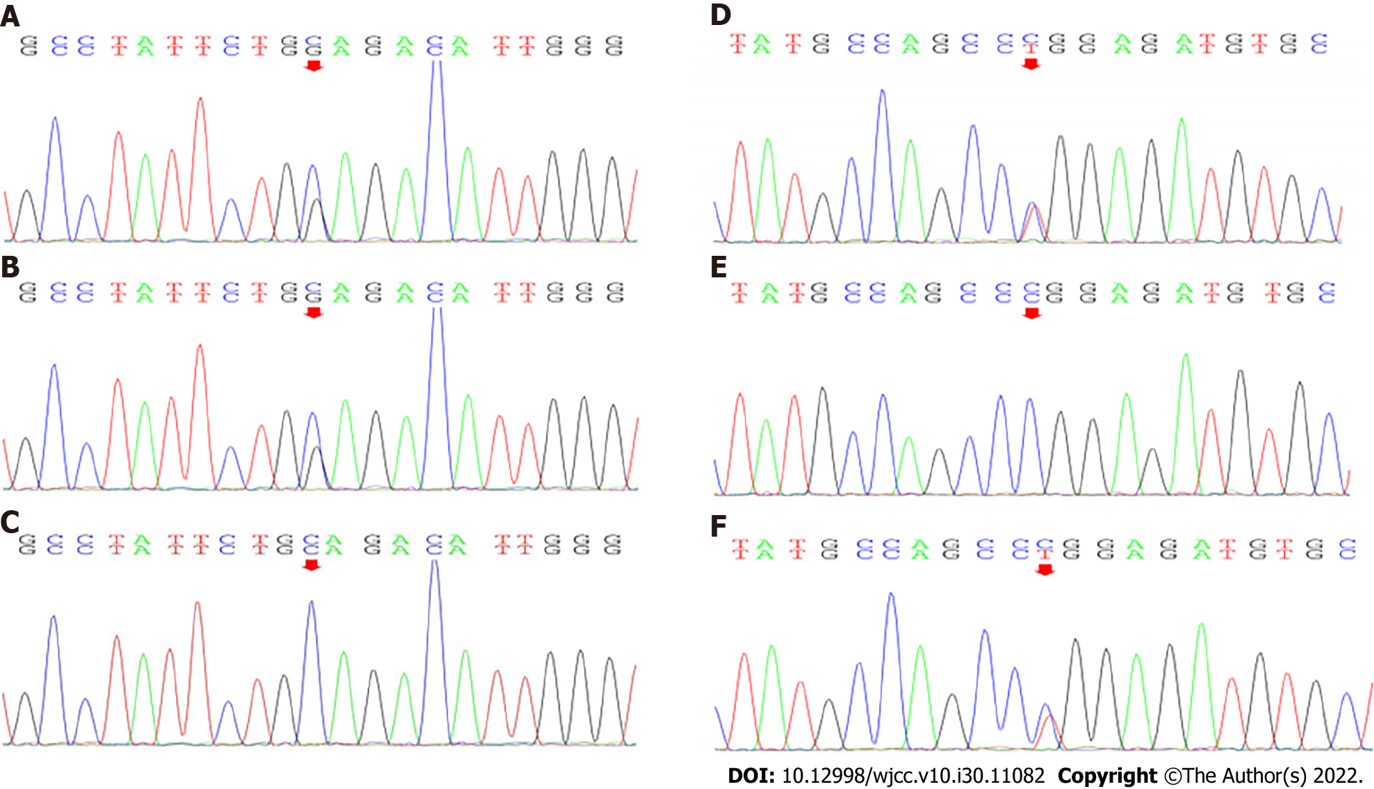
Figure 1 Mutations in the adenylosuccinate lyase genes of the three family members.
A and B: The heterozygous mutation c.154-3C>G was present in the patient and his father; C: The wild-type sequence was present in his mother; D and E: The heterozygous mutation c.71C>T was present in the patient and his mother; F: The wild-type sequence was present in his father.
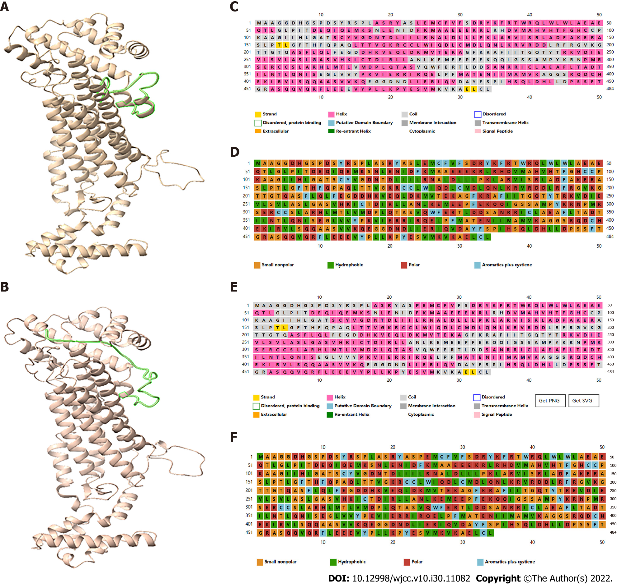
Figure 2 Structures of the wild-type and mutant adenylosuccinate lyase.
A: The tertiary structure of wild-type mutant adenylosuccinate lyase (ADSL); B: The tertiary structure of mutant ADSL; C: The secondary structure of wild-type ADSL; D: The predicted results of amino acid polarity in wild-type ADSL; E: The secondary structure of p. Pro24Leu ADSL; F: The predicted results of amino acid polarity in p. Pro24Leu ADSL. The green regions in panels A and B indicate substantial changes in protein structure.
Figure 3 SD-Score Algorithm predicted that the mutation would affect gene splicing.
Ri: Information contents; CV: Position weight matrix; ΔSD-Score: Differences in the SD-Score; ΔRi: Differences in the information contents; ΔCV: Differences in the position weight matrix.
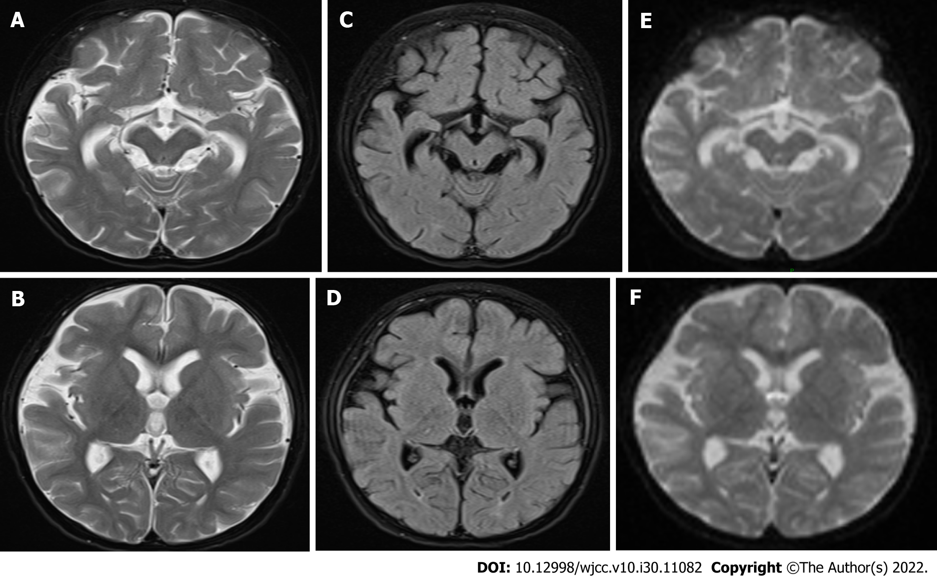
Figure 4 Brain magnetic resonance images findings.
A and B: T2-weighted images; C and D: Diffusion-weighted images; E and F: Fluid-attenuated inversion recovery images. Magnetic resonance images showed bilateral external frontal temporal space widening and abnormal signals around the posterior horns of both lateral ventricles.
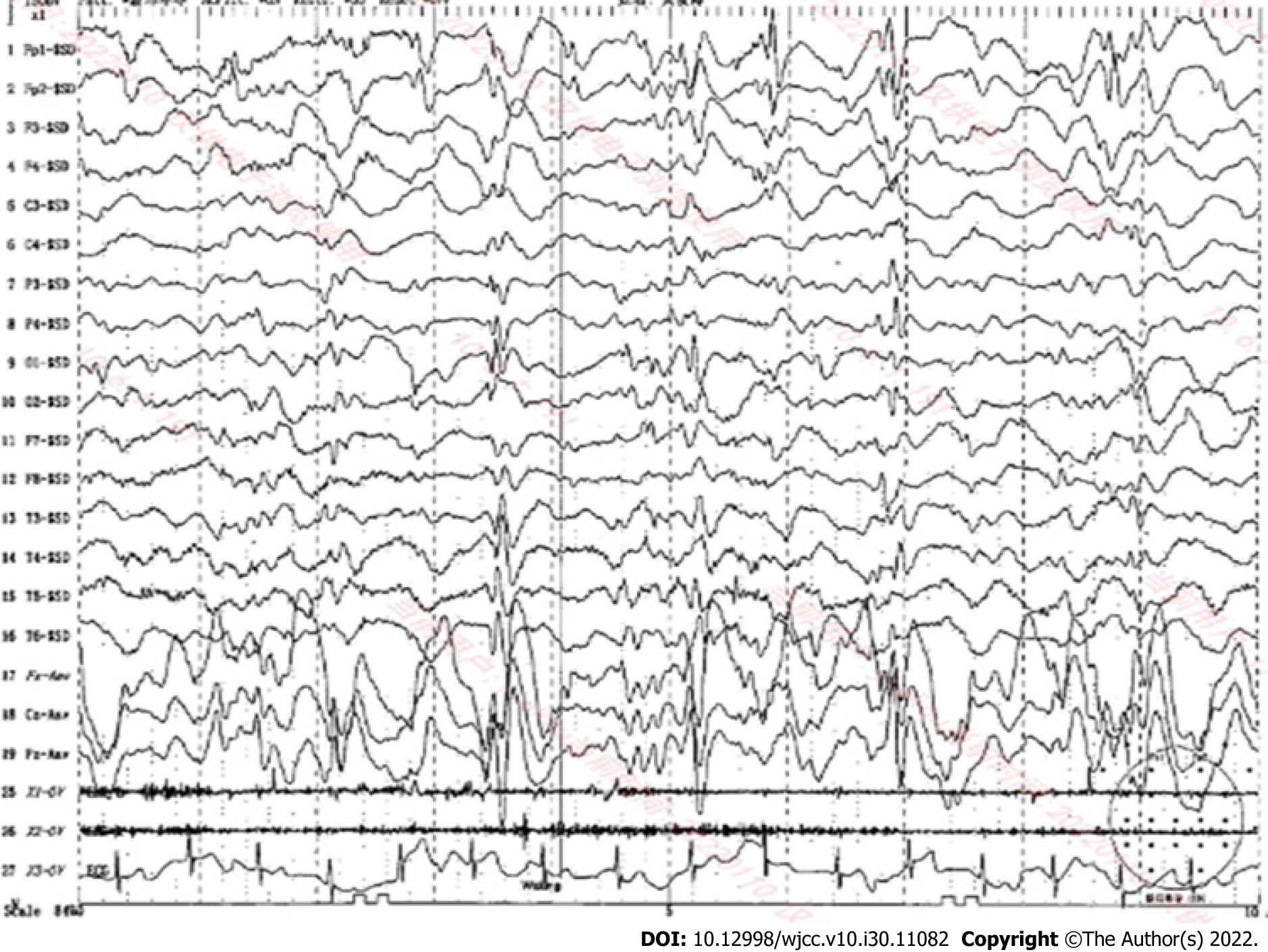
Figure 5
Electroencephalography findings.
- Citation: Wang XC, Wang T, Liu RH, Jiang Y, Chen DD, Wang XY, Kong QX. Child with adenylosuccinate lyase deficiency caused by a novel complex heterozygous mutation in the ADSL gene: A case report. World J Clin Cases 2022; 10(30): 11082-11089
- URL: https://www.wjgnet.com/2307-8960/full/v10/i30/11082.htm
- DOI: https://dx.doi.org/10.12998/wjcc.v10.i30.11082


