Copyright
©The Author(s) 2022.
World J Clin Cases. Oct 26, 2022; 10(30): 11059-11065
Published online Oct 26, 2022. doi: 10.12998/wjcc.v10.i30.11059
Published online Oct 26, 2022. doi: 10.12998/wjcc.v10.i30.11059
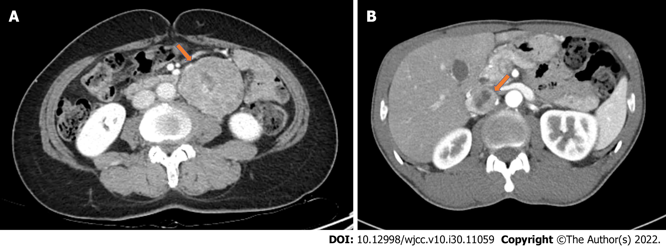
Figure 1 Abdominal computed tomography.
A: Abdominal computed tomography (CT) showing a 6-cm-sized well-circumscribed heterogeneously enhancing mass around duodenojejunal flexure; B: Abdominal CT showing an approximately 3-cm-sized retroperitoneal mass in the retrocaval space with anterior displacement of the inferior vena cava.
- Citation: Kang D, Kim BE, Hong M, Kim J, Jeong S, Lee S. Different intraoperative decisions for undiagnosed paraganglioma: Two case reports. World J Clin Cases 2022; 10(30): 11059-11065
- URL: https://www.wjgnet.com/2307-8960/full/v10/i30/11059.htm
- DOI: https://dx.doi.org/10.12998/wjcc.v10.i30.11059


