Copyright
©The Author(s) 2022.
World J Clin Cases. Oct 26, 2022; 10(30): 11044-11048
Published online Oct 26, 2022. doi: 10.12998/wjcc.v10.i30.11044
Published online Oct 26, 2022. doi: 10.12998/wjcc.v10.i30.11044
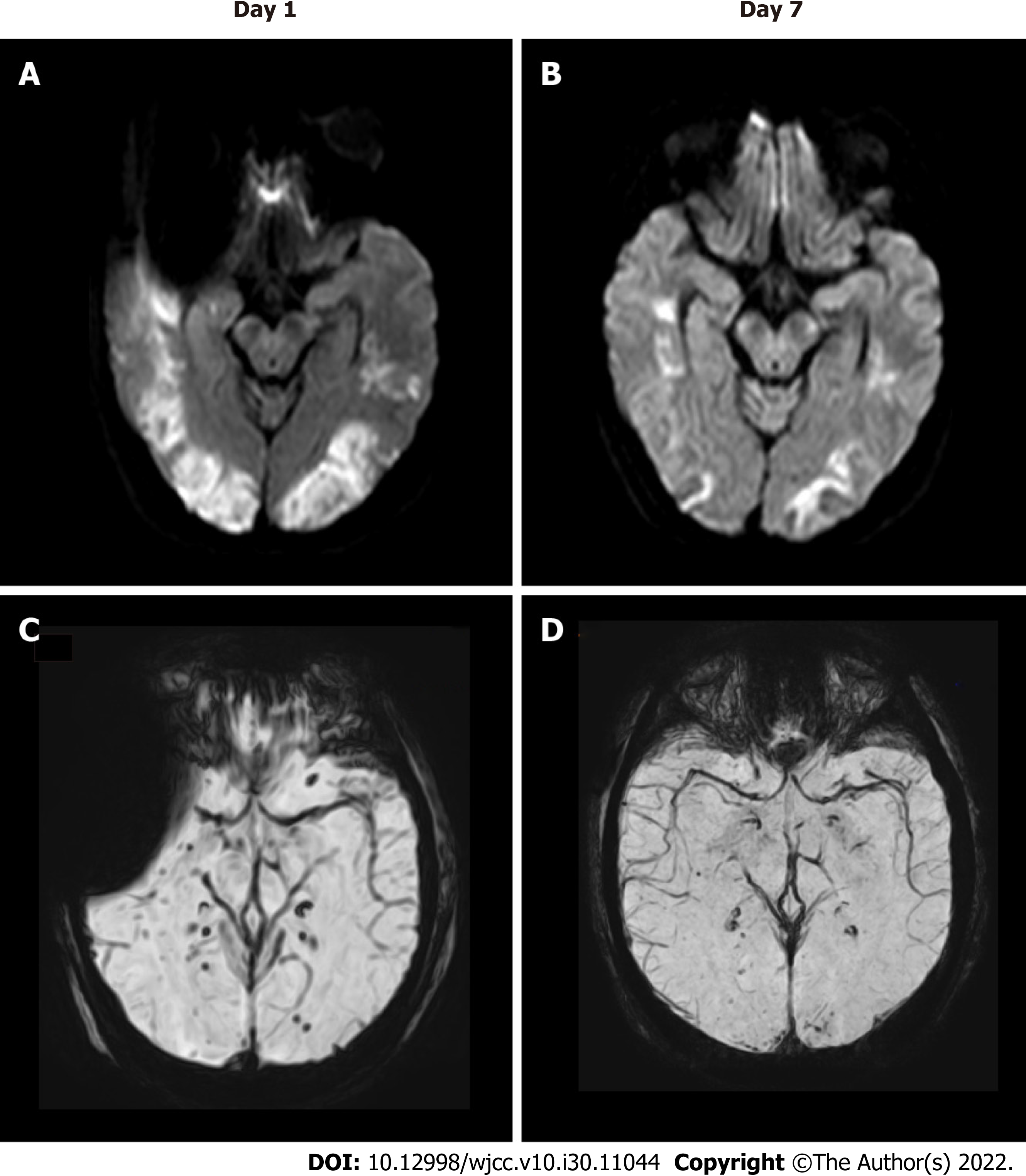
Figure 1 Brain magnetic resonance imaging showed bilateral multifocal vasogenic edema especially in bilateral occipital lobes.
A: Compatible with posterior reversible encephalopathy syndrome; B and D: Follow-up magnetic resonance imaging after 7 d showed significantly reduced vasogenic edema in both cerebral hemispheres, with decreased microbleeds on susceptibility weighted imaging (SWI) mapping; C: Spot-like microbleeds were found on SWI mapping.
- Citation: Dai SJ, Yu QJ, Zhu XY, Shang QZ, Qu JB, Ai QL. Autoimmune encephalitis with posterior reversible encephalopathy syndrome: A case report. World J Clin Cases 2022; 10(30): 11044-11048
- URL: https://www.wjgnet.com/2307-8960/full/v10/i30/11044.htm
- DOI: https://dx.doi.org/10.12998/wjcc.v10.i30.11044


