Copyright
©The Author(s) 2022.
World J Clin Cases. Jan 21, 2022; 10(3): 1050-1055
Published online Jan 21, 2022. doi: 10.12998/wjcc.v10.i3.1050
Published online Jan 21, 2022. doi: 10.12998/wjcc.v10.i3.1050
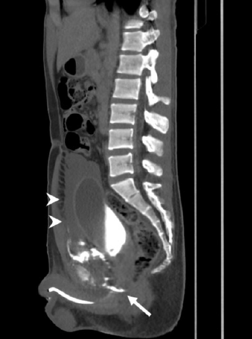
Figure 1 Contrast medium extravasation at bulbar urethra (arrow) with massive hematoma (arrow heads), and “pie in the sky” sign of prostate was noted.
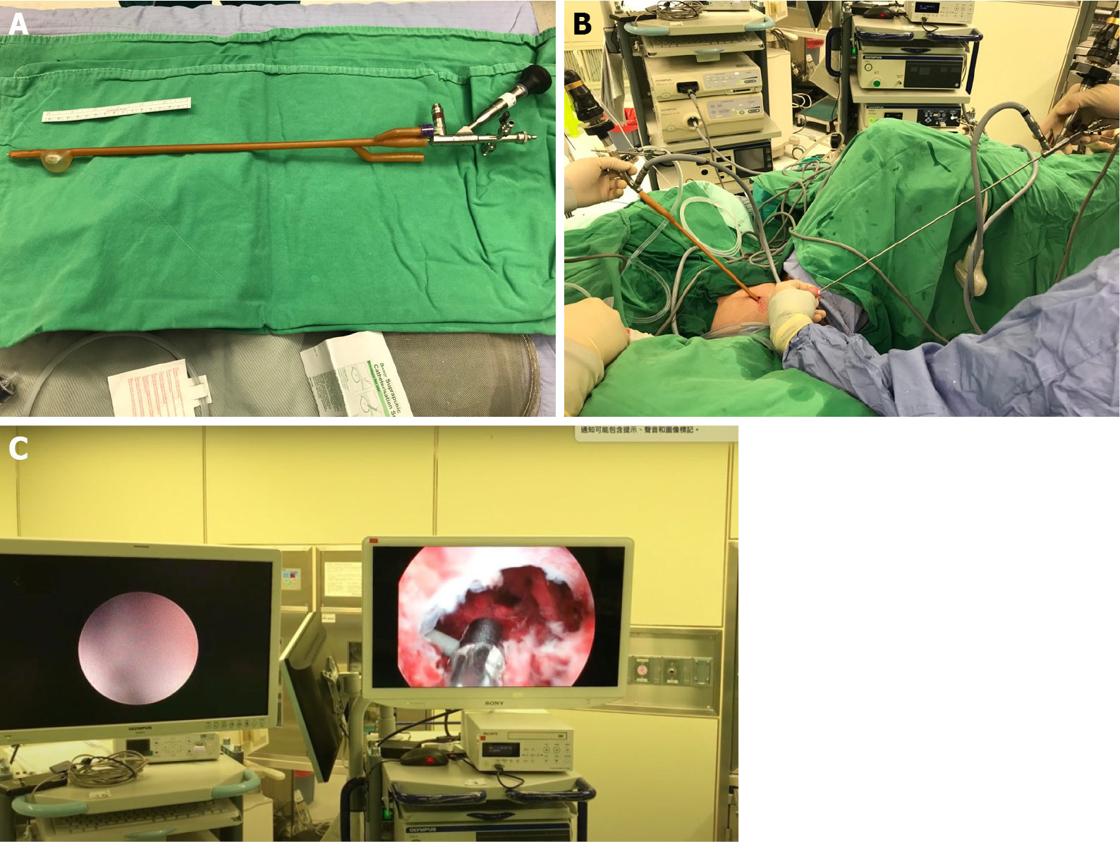
Figure 2 Procedure of primary endoscopic realignment.
A: The tip of the Foley catheter was incised and a semi-rigid 4.5/6.5-Fr ureteroscope was inserted; B: Simultaneous antegrade and retrograde endoscopy; C: The guidewire was antegrade and then pulled out through the external urethral meatus using grasping forceps with a cystoscope.
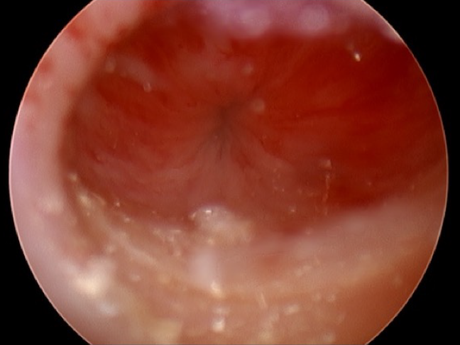
Figure 3 Healed urethra at 28 d after realignment.
- Citation: Ho CJ, Yang MH. Novel method of primary endoscopic realignment for high-grade posterior urethral injuries: A case report. World J Clin Cases 2022; 10(3): 1050-1055
- URL: https://www.wjgnet.com/2307-8960/full/v10/i3/1050.htm
- DOI: https://dx.doi.org/10.12998/wjcc.v10.i3.1050


