Copyright
©The Author(s) 2022.
World J Clin Cases. Sep 26, 2022; 10(27): 9805-9813
Published online Sep 26, 2022. doi: 10.12998/wjcc.v10.i27.9805
Published online Sep 26, 2022. doi: 10.12998/wjcc.v10.i27.9805
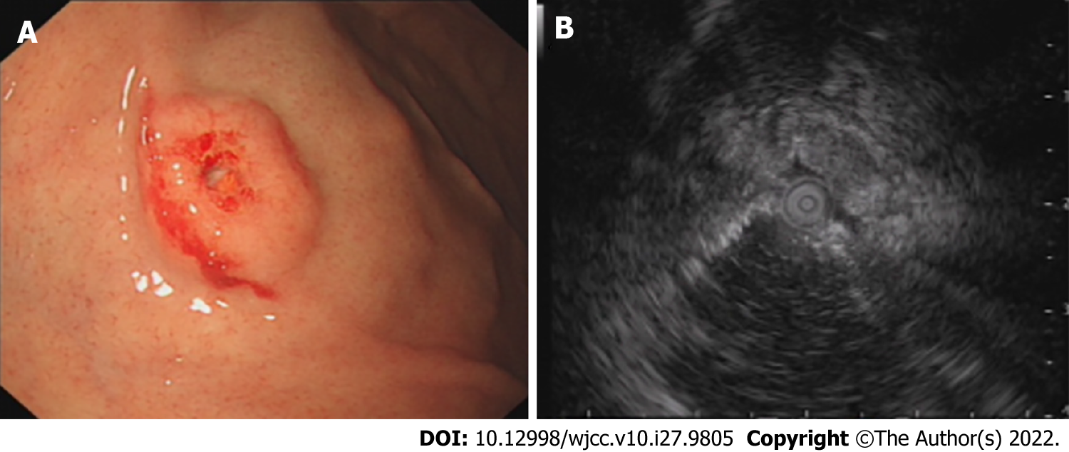
Figure 1 Endoscopic ultrasonography examination in the local hospital 1 year previously.
A: A solitary discoid-shaped submucosal tumor in the gastric fundus with central depression and surface mucosal congestion (IIa+IIc appearance, moderately irregular edges, presence of 2 mucosal folds converging on the lesion); B: The lesion originated from the deeper layers of the mucosa with partially discontinuous submucosa, medium and hypoechoic changes, and was 1.12 cm × 0.38 cm in size.
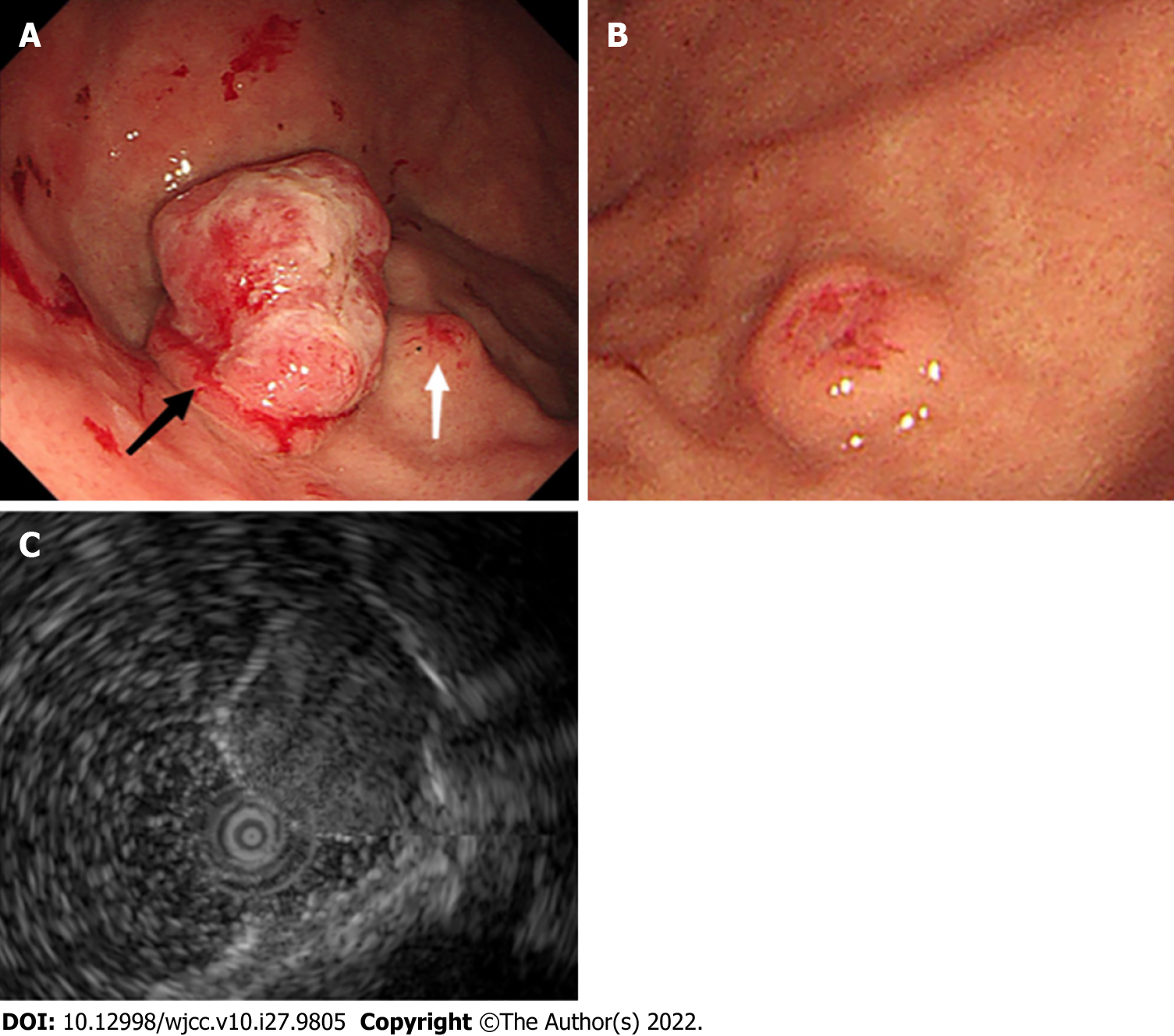
Figure 2 Endoscopic ultrasonography examination in our hospital.
A: A large protruding lesion was found in the fundus, the primary discoid-shape remained in the basal layer of the lesion (black arrow). A small submucosal lesion was detected in the fundus adjacent to the large lesion (white arrow); B: The other similar small submucosal lesion in the middle section of the stomach body; and C: EUS showed a heterogeneous mass that involved the mucosa and submucosal layer, with hypoechoic changes.
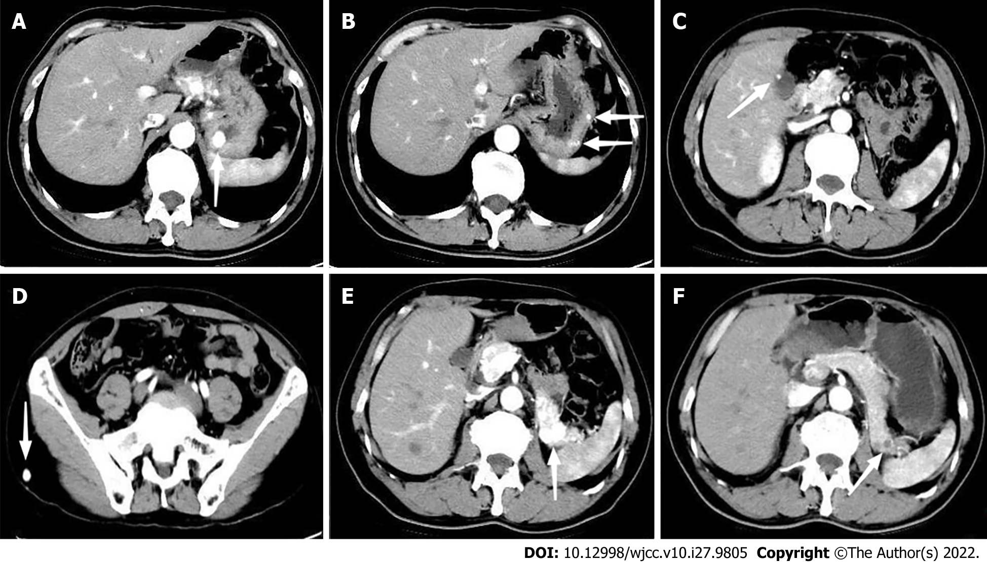
Figure 3 Abdominal computed tomography revealed multiple hypervascular lesions with abnormal enhancement in the arterial phases.
A: Lesion in the fundus; B: Multiple lesions in the body of the stomach; C: Lesion in the gallbladder; D: Lesion in the subcutaneous soft tissue of the right buttock; E: Multiple lesions in the pancreas. F: The lesions in the pancreas became smaller, and enhancement in the arterial phase reduced during the 8-mo follow-up period after axitinib therapy.
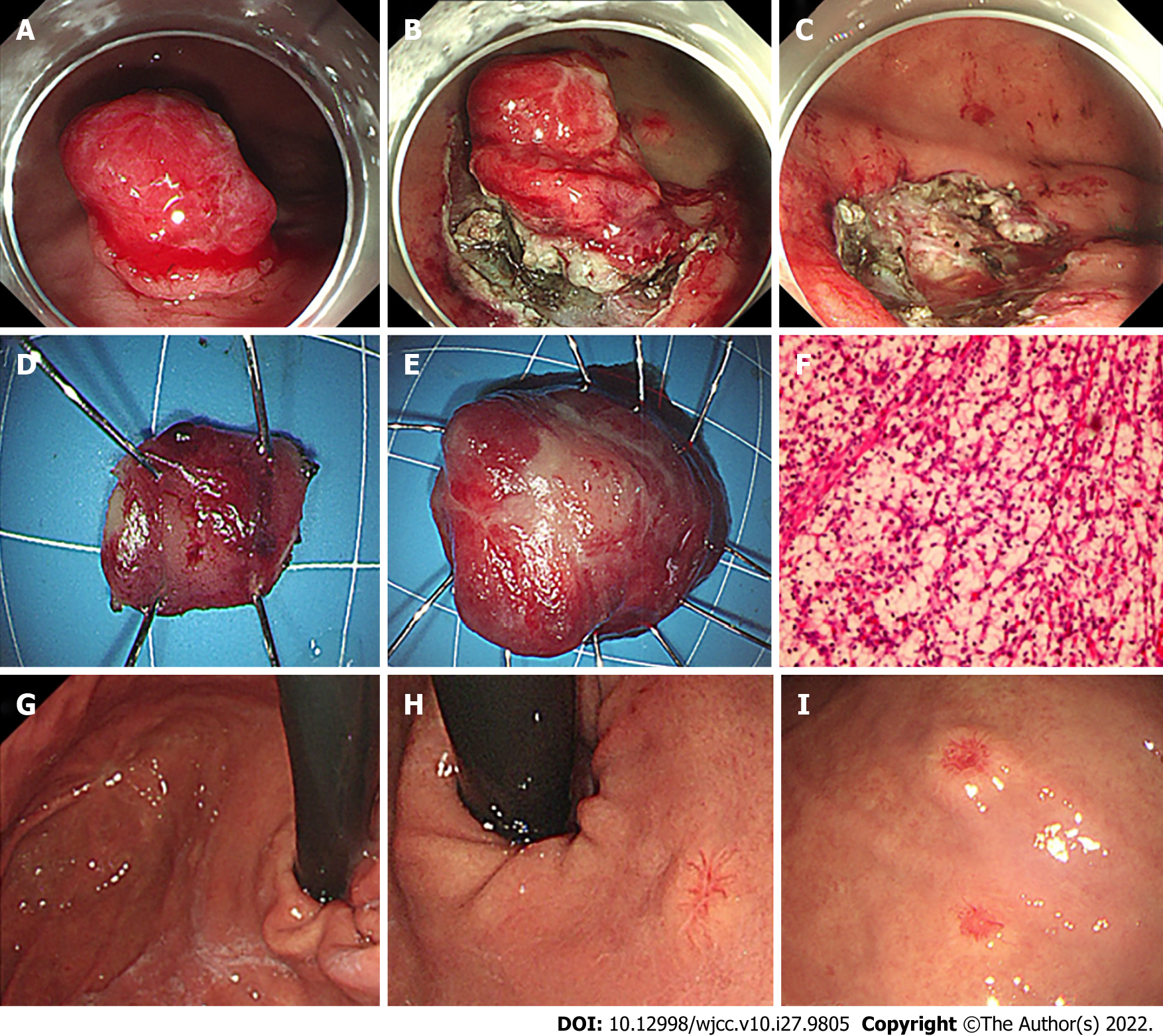
Figure 4 Our endoscopic management and 1-year follow-up.
A-E: Two adjacent lesions were resected endoscopically by endoscopic submucosal dissection; F: The postoperative pathological analysis confirmed gastric metastasis from clear cell RCC; G: At 1-year follow-up, gastroscopy examination showed scars in the fundus with no relapse; H: Two new small lesions emerged, one in the upper section of the stomach body close to the cardia; and I: The other in the stomach body adjacent to the previous lesion.
- Citation: Chen WG, Shan GD, Zhu HT, Chen LH, Xu GQ. Gastric metastasis presenting as submucosa tumors from renal cell carcinoma: A case report. World J Clin Cases 2022; 10(27): 9805-9813
- URL: https://www.wjgnet.com/2307-8960/full/v10/i27/9805.htm
- DOI: https://dx.doi.org/10.12998/wjcc.v10.i27.9805


