Copyright
©The Author(s) 2022.
World J Clin Cases. Jul 6, 2022; 10(19): 6437-6445
Published online Jul 6, 2022. doi: 10.12998/wjcc.v10.i19.6437
Published online Jul 6, 2022. doi: 10.12998/wjcc.v10.i19.6437
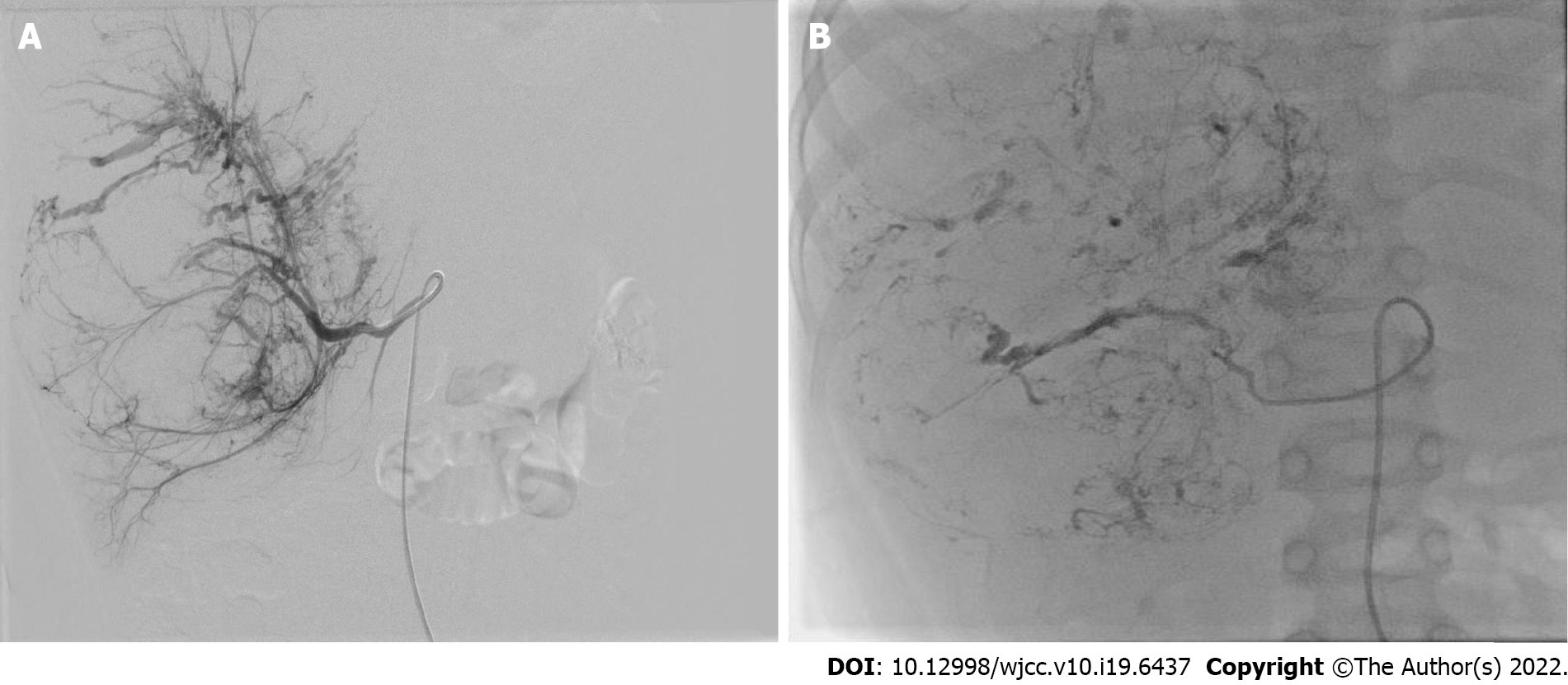
Figure 1 Transcatheter arterial chemoembolization procedure of case 6.
A: The catheter was introduced into the tumor-feeding artery and performed angiography, showing the stereoscopic configuration of "holding ball"; B: After injection of anticarcinogen and lipiodol, the outline of the entire tumor was revealed.
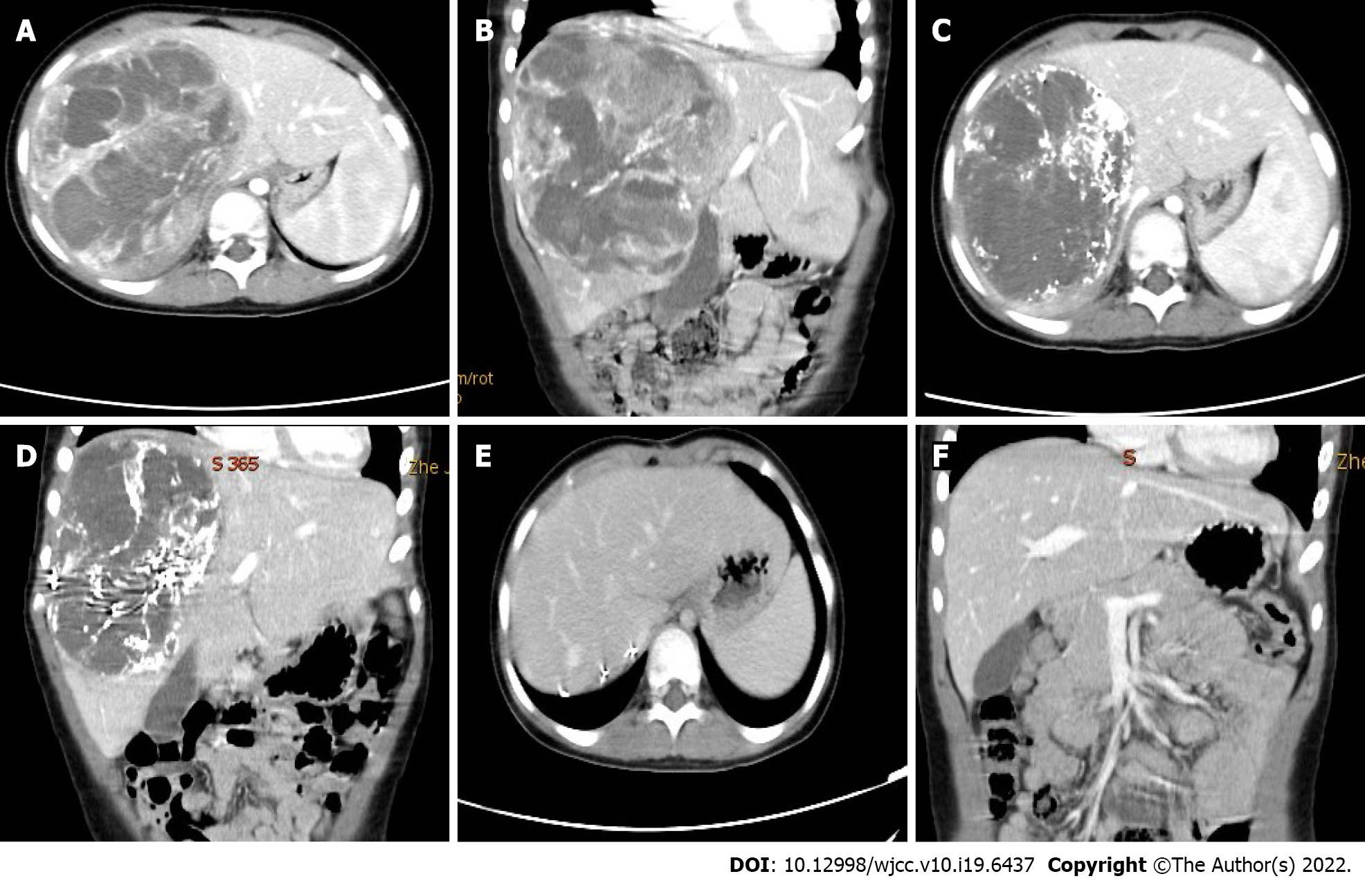
Figure 2 The imaging manifestation of case 6.
A and B: Computed tomography scans showed a giant mass of the right liver (diameter 14.8 cm); C and D: One month after neoadjuvant therapy: The tumor volumes shrunk by 48.8%, lipiodol deposits in the tumor with a clear boundary; E and F: One month after tumor resection.
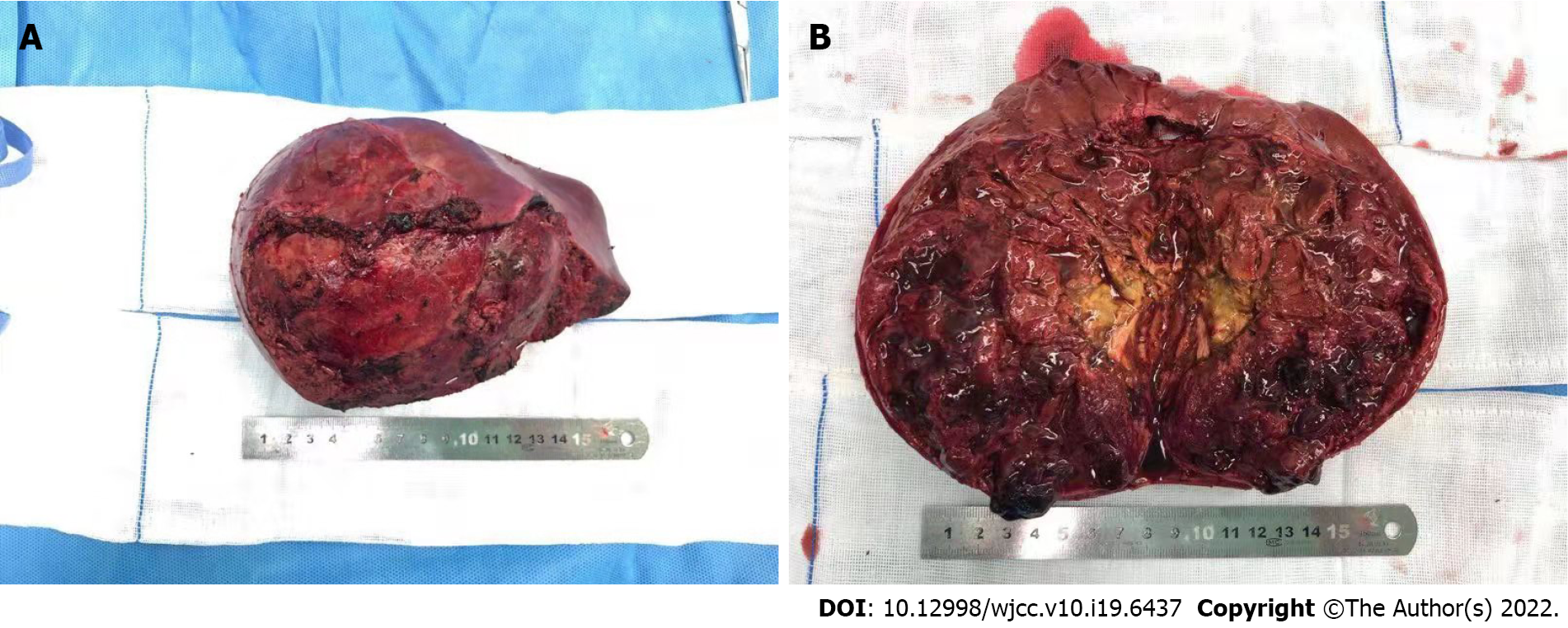
Figure 3 These are conducive to complete resection, reducing the rate of positive surgical margins, and diminishing intraoperative bleeding.
A: The gross of tumor appears as border clear with about 10 cm in diameter; B: The cut surface is red, brown or yellow and has soft qualitative with focal necrosis.
- Citation: He M, Cai JB, Lai C, Mao JQ, Xiong JN, Guan ZH, Li LJ, Shu Q, Ying MD, Wang JH. Neoadjuvant transcatheter arterial chemoembolization and systemic chemotherapy for the treatment of undifferentiated embryonal sarcoma of the liver in children. World J Clin Cases 2022; 10(19): 6437-6445
- URL: https://www.wjgnet.com/2307-8960/full/v10/i19/6437.htm
- DOI: https://dx.doi.org/10.12998/wjcc.v10.i19.6437


