Copyright
©The Author(s) 2022.
World J Clin Cases. Jun 26, 2022; 10(18): 6184-6191
Published online Jun 26, 2022. doi: 10.12998/wjcc.v10.i18.6184
Published online Jun 26, 2022. doi: 10.12998/wjcc.v10.i18.6184
Figure 1 Histology and immunohistochemistry images.
A: Histology; B: Positivity for programmed death-ligand 1 (combined positive score = 1); C: Positivity for human epidermal growth factor receptor 2.
Figure 2 Computed tomography images.
A: Baseline; B: After neo-adjuvant therapy; C: After surgery.
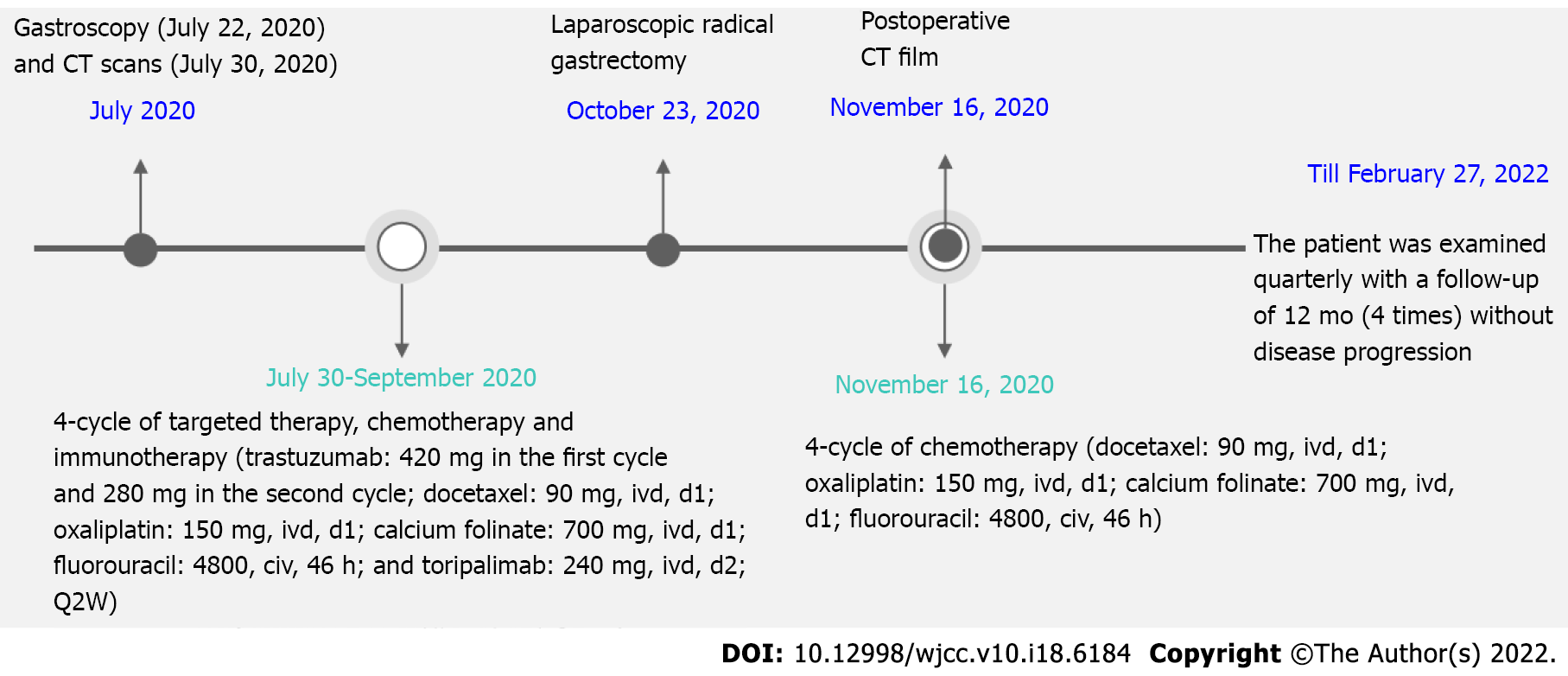
Figure 3 Timeline of this case report.
CT: Computed tomography.
- Citation: Liu R, Wang X, Ji Z, Deng T, Li HL, Zhang YH, Yang YC, Ge SH, Zhang L, Bai M, Ning T, Ba Y. Toripalimab combined with targeted therapy and chemotherapy achieves pathologic complete response in gastric carcinoma: A case report. World J Clin Cases 2022; 10(18): 6184-6191
- URL: https://www.wjgnet.com/2307-8960/full/v10/i18/6184.htm
- DOI: https://dx.doi.org/10.12998/wjcc.v10.i18.6184


