Copyright
©The Author(s) 2022.
World J Clin Cases. Jun 6, 2022; 10(16): 5479-5486
Published online Jun 6, 2022. doi: 10.12998/wjcc.v10.i16.5479
Published online Jun 6, 2022. doi: 10.12998/wjcc.v10.i16.5479
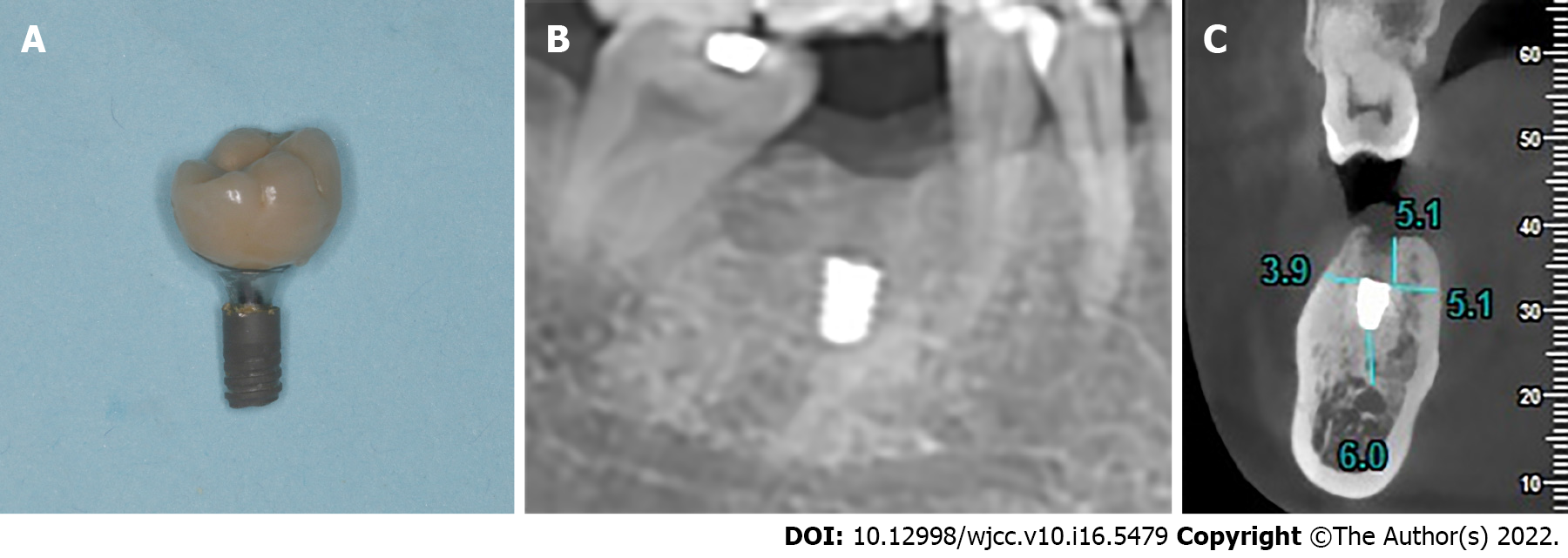
Figure 1 The condition of the fractured implant when the patient came to the clinic.
A: Fractured portion of the implant connected to the prosthesis; B: Panoramic radiograph showed no signs of peri-implantitis; C: CBCT showed that the remaining implant was 6.0 mm away from the inferior alveolar nerve canal, 3.9 mm away from the buccal cortical bone wall, 5.1 mm away from the lingual bone wall and 5.1 mm beneath the crestal ridge.
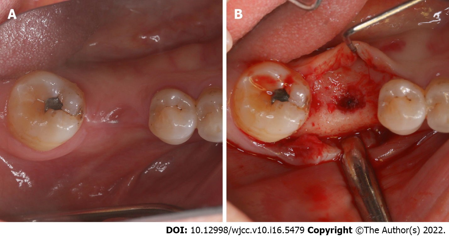
Figure 2 Occlusal view before remaining implant removal.
A: The rapidly growing gingiva had closed the gingival outlet of the implant; B: After a crestal full-thickness flap was raised, the implant hole filled with granulation tissue was observed.
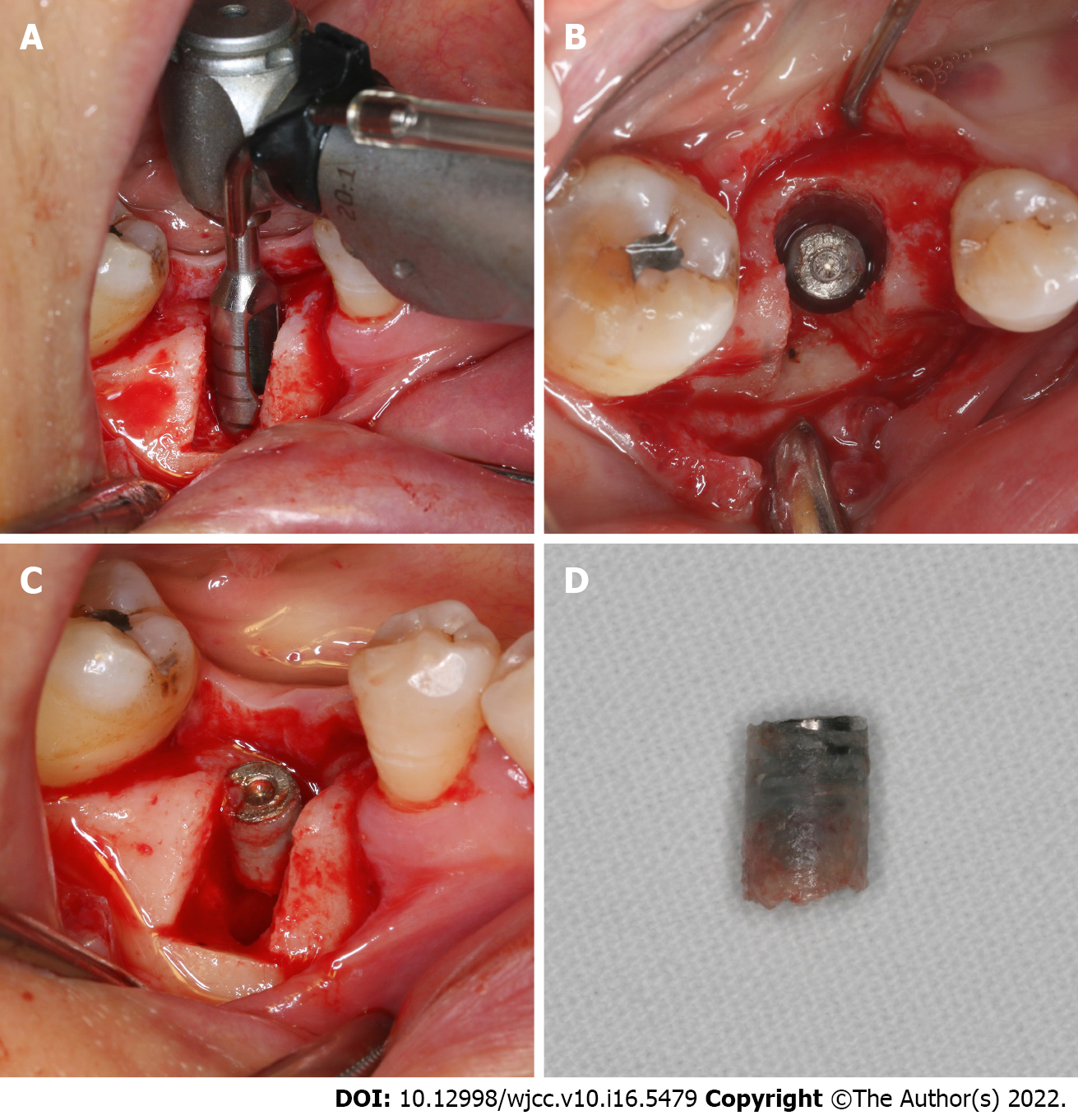
Figure 3 The osteotomy procedure.
A: Two vertical incisions and one horizontal incision were made using an ultrasonic osteotome on the buccal plate; B: The buccal bone plate was removed with a bone chisel and a hammer; C: The broken end of the fractured implant was clearly exposed.
Figure 4 Removal of the fractured implant with a trephine.
A: The remaining implant was completely removed with a graduated trephine; B: A ring of uniform thickness was created around the remaining implant from the occlusal view; C: The remaining segment was moved out; D: The surface of the implant was covered with a thin layer of osseointegrated alveolar bone.
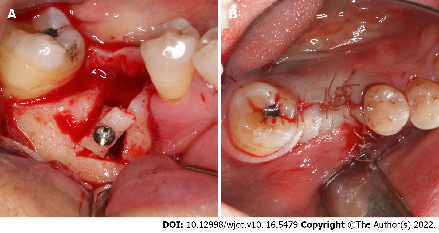
Figure 5 A modified Wafer technique-supported guided bone regeneration treatment was conducted simultaneously to preserve the horizontal alveolar dimension.
A: The buccal bone plate was repositioned in situ using a titanium nail with slight rotation; B: The wound was closed up tightly.
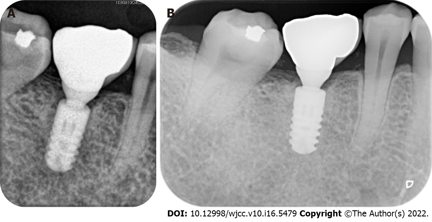
Figure 6 The periapical radiographic examination.
A: Immediately after the crown restoration showed a well-osseointegrated new implant; B: After 12 mo of function.
- Citation: Chen LW, Wang M, Xia HB, Chen D. Osteotomy combined with the trephine technique for invisible implant fracture: A case report. World J Clin Cases 2022; 10(16): 5479-5486
- URL: https://www.wjgnet.com/2307-8960/full/v10/i16/5479.htm
- DOI: https://dx.doi.org/10.12998/wjcc.v10.i16.5479


