Copyright
©The Author(s) 2022.
World J Clin Cases. Jun 6, 2022; 10(16): 5365-5372
Published online Jun 6, 2022. doi: 10.12998/wjcc.v10.i16.5365
Published online Jun 6, 2022. doi: 10.12998/wjcc.v10.i16.5365
Figure 1 Computed tomography scan of the head on day 1 of life.
A and B revealed massive brain edema and left parenchymal brain hemorrhage with extension into ventricular and subarachnoid spaces.
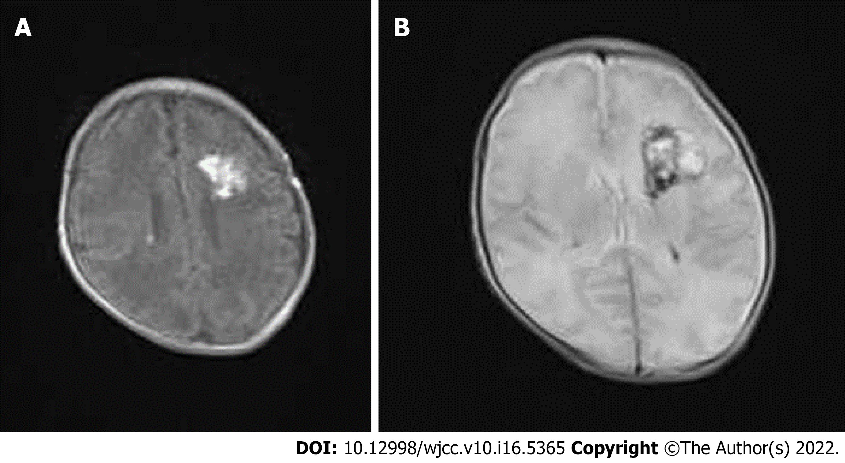
Figure 2 Magnetic resonance imaging of the patient on day 25 of life.
Follow-up magnetic resonance imaging of the patient on day 25 of life revealed improvement of left parenchymal hemorrhage and subarachnoid hemorrhage. A: Hypersignal on T1; B: Distinctive heterogeneous signal on T2.
- Citation: Lu Y, Zhang ZQ. Neonatal hemorrhage stroke and severe coagulopathy in a late preterm infant after receiving umbilical cord milking: A case report. World J Clin Cases 2022; 10(16): 5365-5372
- URL: https://www.wjgnet.com/2307-8960/full/v10/i16/5365.htm
- DOI: https://dx.doi.org/10.12998/wjcc.v10.i16.5365


