Copyright
©The Author(s) 2022.
World J Clin Cases. Jun 6, 2022; 10(16): 5331-5336
Published online Jun 6, 2022. doi: 10.12998/wjcc.v10.i16.5331
Published online Jun 6, 2022. doi: 10.12998/wjcc.v10.i16.5331
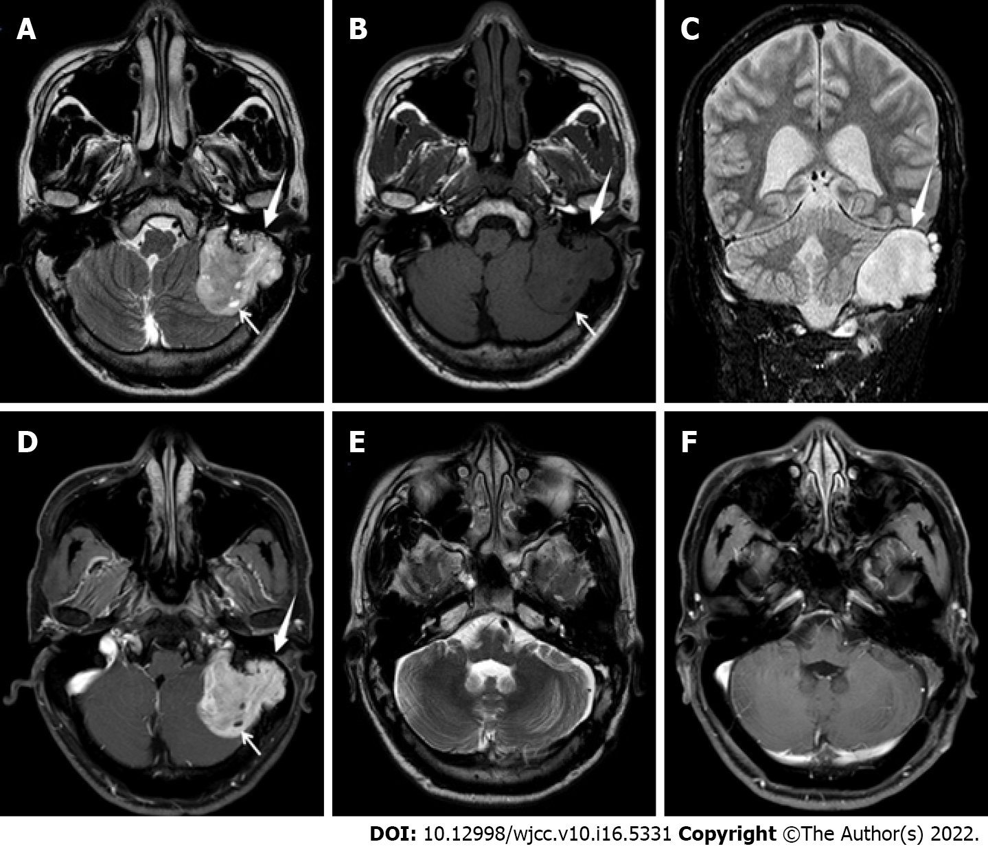
Figure 1 Brain magnetic resonance imaging before surgery.
A, B: T2-weighted axial images (A) and T1-weighted images (B) showed a lobulated mass (white thick arrow) in the petrous part and mastoid area of the left temporal bone, swelling to the brain, with isointense and hypointensity signal on T1-weighted images, hyperintensity and slightly hyperintensity signal on T2-weighted images , and there was a small cystic degeneration area inside (white fine arrow), the surrounding bone was damaged, and the left cerebellar hemisphere was compressed to deformation; C: Fluid-attenuated inversion recovery coronal magnetic resonance imaging (MRI) showed a mass with hyperintensity signal (white thick arrow), and the boundary was clear; D: T1-enhanced axial sequence showed uneven obvious contrast enhancement. There was no enhancement in the cystic degeneration area (white small arrow). Brain MRI at 5 years after surgery; E, F: T2-weighted axial images (E) and T1-weighted images enhanced (F) sequence showed postoperative changes in the left mastoid area, but no definite abnormal signs were found.
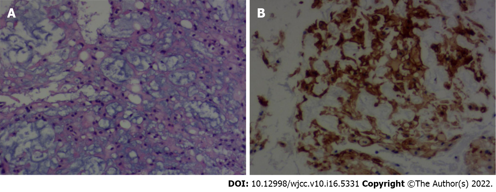
Figure 2 Histological examination of chordoma.
A: Epithelioid polygonal cells with clear or palely eosinophilic cytoplasm arranged in clusters and cords (hematoxylin and eosin, 100×). B: Immunohistochemical examination showed positive expression of epithelial membrane antigen (magnification 100×).
- Citation: Hua JJ, Ying ML, Chen ZW, Huang C, Zheng CS, Wang YJ. Chordoma of petrosal mastoid region: A case report. World J Clin Cases 2022; 10(16): 5331-5336
- URL: https://www.wjgnet.com/2307-8960/full/v10/i16/5331.htm
- DOI: https://dx.doi.org/10.12998/wjcc.v10.i16.5331


