Copyright
©The Author(s) 2022.
World J Clin Cases. May 16, 2022; 10(14): 4569-4573
Published online May 16, 2022. doi: 10.12998/wjcc.v10.i14.4569
Published online May 16, 2022. doi: 10.12998/wjcc.v10.i14.4569
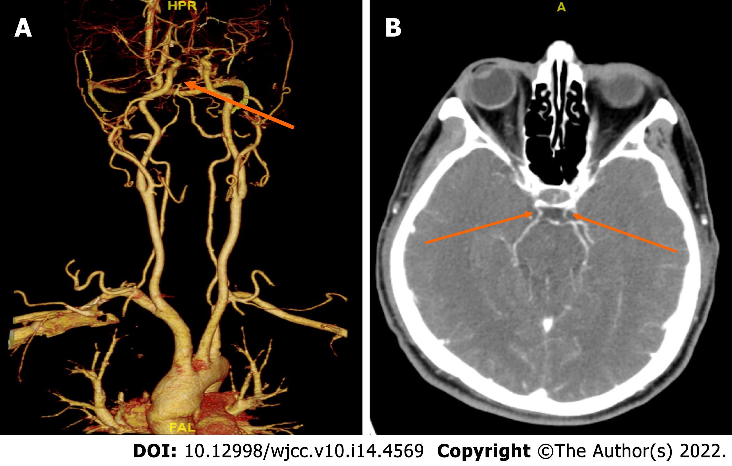
Figure 1 Computed tomography angiography imaging.
A: Basilar artery occlusion; B: Posterior communicating artery was patent.
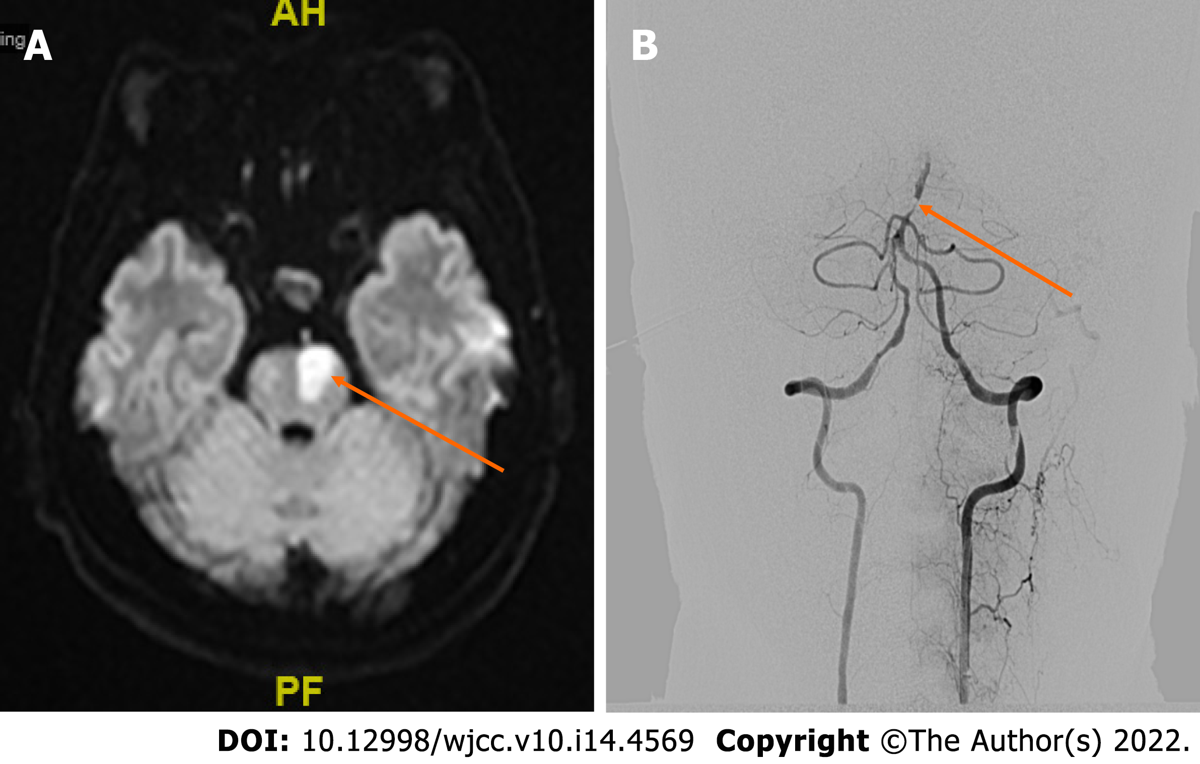
Figure 2 Further diagnostic work-up.
A: Magnetic resonance diffusion weighted imaging suggests left pontine infarction; B: Digital subtraction angiography shows severe stenosis in the lower segment of the basilar artery and occlusion in the upper segment.
- Citation: Wang TL, Wu G, Liu SZ. Convulsive-like movements as the first symptom of basilar artery occlusive brainstem infarction: A case report. World J Clin Cases 2022; 10(14): 4569-4573
- URL: https://www.wjgnet.com/2307-8960/full/v10/i14/4569.htm
- DOI: https://dx.doi.org/10.12998/wjcc.v10.i14.4569


