Copyright
©The Author(s) 2022.
World J Clin Cases. May 6, 2022; 10(13): 4196-4206
Published online May 6, 2022. doi: 10.12998/wjcc.v10.i13.4196
Published online May 6, 2022. doi: 10.12998/wjcc.v10.i13.4196
Figure 1 Contrast-enhanced chest computed tomography images of (A, B) unenhanced and (C) enhanced scan.
A 6.9-cm diameter well-circumscribed mass in the left lower lobe of the lung shows mild homogeneous enhancement.
Figure 2 Positive uptake by the mass on 18F-fluorodeoxyglucose-positron emission tomography suggesting malignancy.
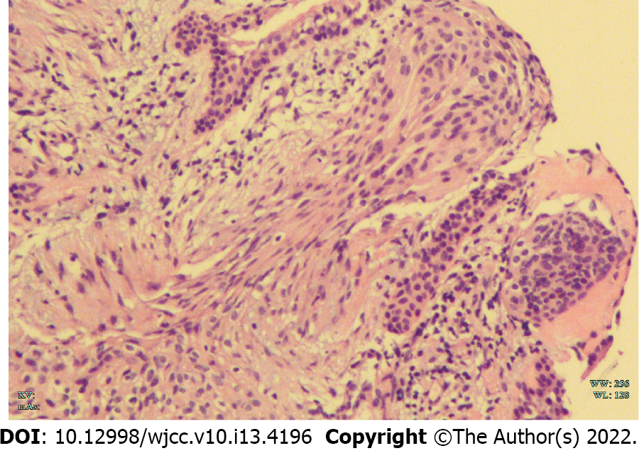
Figure 3 The transbronchial biopsy result: Hematoxylin and eosin staining showed that a few nested epithelioid cells and abnormal cells were observed in the tissue (200×).
Figure 4 Histological features of primary pulmonary meningioma.
A-D: Macroscopically, primary pulmonary meningioma (PPM) showed as spindle or oval cells organized in bundles and whorls on hematoxylin-eosin staining (25×; 50×; 100×; 200×); E-H: Immunohistochemically (200×), PPM showed negativity for E: Cytokeratin, positive for F: Epithelial membrane antigen; G: Progesterone receptor; H: Somatostatin Receptor 2 (SSTR2).
- Citation: Zhang DB, Chen T. Primary pulmonary meningioma: A case report and review of the literature. World J Clin Cases 2022; 10(13): 4196-4206
- URL: https://www.wjgnet.com/2307-8960/full/v10/i13/4196.htm
- DOI: https://dx.doi.org/10.12998/wjcc.v10.i13.4196


