
















©The Author(s) 2017.
World J Nephrol. Sep 6, 2017; 6(5): 221-228
Published online Sep 6, 2017. doi: 10.5527/wjn.v6.i5.221
Published online Sep 6, 2017. doi: 10.5527/wjn.v6.i5.221
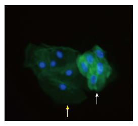
Figure 2 A cluster of podocytes diagnosed by immunofluorescence in a patient with Fabry disease are shown to the right (white arrow).
Synaptopodin has been employed as the targeted podocyte protein. Tubular cells are also seen to the left of the image (yellow arrow). Green color stands for cytoplasmic synaptopodin, while blue color discloses cellular nuclei × 400.
- Citation: Trimarchi H. Podocyturia: Potential applications and current limitations. World J Nephrol 2017; 6(5): 221-228
- URL: https://www.wjgnet.com/2220-6124/full/v6/i5/221.htm
- DOI: https://dx.doi.org/10.5527/wjn.v6.i5.221