
















©The Author(s) 2017.
World J Nephrol. Sep 6, 2017; 6(5): 221-228
Published online Sep 6, 2017. doi: 10.5527/wjn.v6.i5.221
Published online Sep 6, 2017. doi: 10.5527/wjn.v6.i5.221
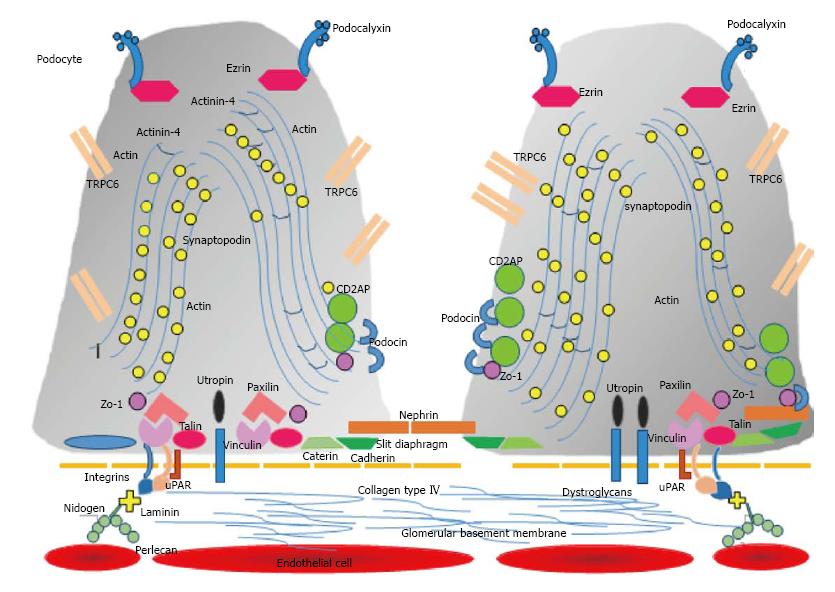
Figure 1 Glomerular filtration barrier.
The podocyte with two of its many foot processes are attached to the glomerular basement membrane forming the slit diaphragm. Some components of these structures are illustrated. The fenestrated endothelium is depicted next to the basement membrane. TRPC6: Transient receptor potential cation channel 6; uPAR: Urokinase-type plasminogen activator receptor.
- Citation: Trimarchi H. Podocyturia: Potential applications and current limitations. World J Nephrol 2017; 6(5): 221-228
- URL: https://www.wjgnet.com/2220-6124/full/v6/i5/221.htm
- DOI: https://dx.doi.org/10.5527/wjn.v6.i5.221