Copyright
©The Author(s) 2016.
World J Nephrol. Sep 6, 2016; 5(5): 461-470
Published online Sep 6, 2016. doi: 10.5527/wjn.v5.i5.461
Published online Sep 6, 2016. doi: 10.5527/wjn.v5.i5.461
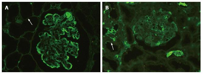
Figure 1 Examples of technically adequate and inadequate digestion.
A: Immunofluorescence staining on a paraffin embedded tissue section in a case of diffuse proliferative lupus nephritis after enzymatic digestion with proteinase K. Note the adequate digestion evidenced by disappearance of tubular epithelial cells (arrow) (FITC IgG, × 200); B: Immunofluorescence staining in a case with inadequate digestion with visible tubular epithelial cells (arrow). Note the antibody sticking to the surface of the capillary wall (FITC IgG, × 200). FITC: Fluorescein isothiocyanate.
- Citation: Singh G, Singh L, Ghosh R, Nath D, Dinda AK. Immunofluorescence on paraffin embedded renal biopsies: Experience of a tertiary care center with review of literature. World J Nephrol 2016; 5(5): 461-470
- URL: https://www.wjgnet.com/2220-6124/full/v5/i5/461.htm
- DOI: https://dx.doi.org/10.5527/wjn.v5.i5.461


