Copyright
©2013 Baishideng.
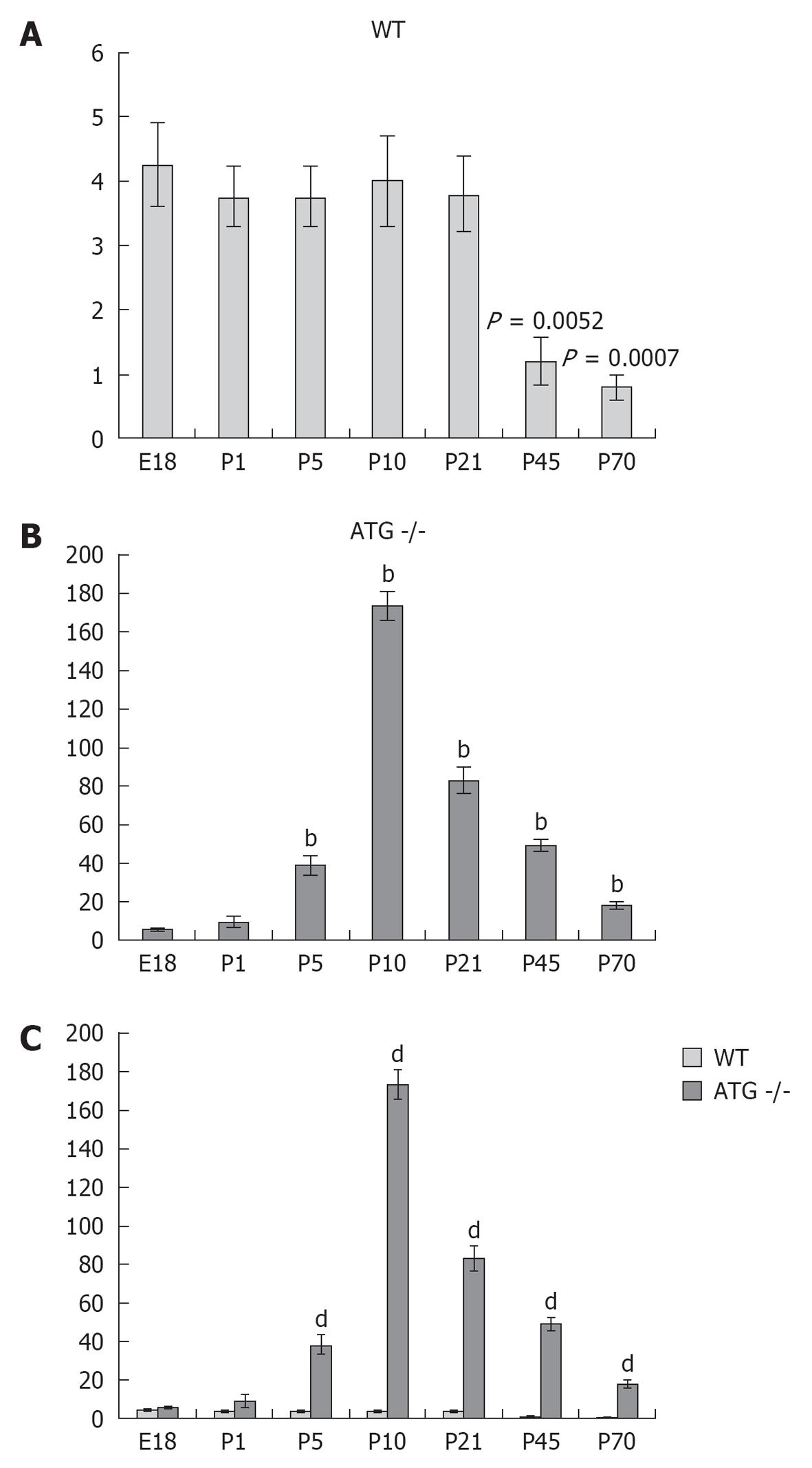
Figure 3 Quantification and comparison of pericytes positive for renin in wild type and angiotensinogen deficient mice.
A: In wild type (WT) mice the density of renin positive pericytes was not significantly different from E18 to P21, but significantly decreased at P45 (P = 0.0052 vs E18) and P70 (P = 0.0007 vs E18); B: Angiotensinogen deficient (AGT) -/- mice showed a significant increase at P5 with a maximum at P10 and a progressive decrease towards P70 (bP≤ 0.01 vs E18); C: Comparison at each individual age showing a significant increase in the number of renin-positive pericytes in AGT -/- from P5 till P70 (dP≤ 0.001 vs WT).
- Citation: Berg AC, Chernavvsky-Sequeira C, Lindsey J, Gomez RA, Sequeira-Lopez MLS. Pericytes synthesize renin. World J Nephrol 2013; 2(1): 11-16
- URL: https://www.wjgnet.com/2220-6124/full/v2/i1/11.htm
- DOI: https://dx.doi.org/10.5527/wjn.v2.i1.11


