Copyright
©2013 Baishideng.
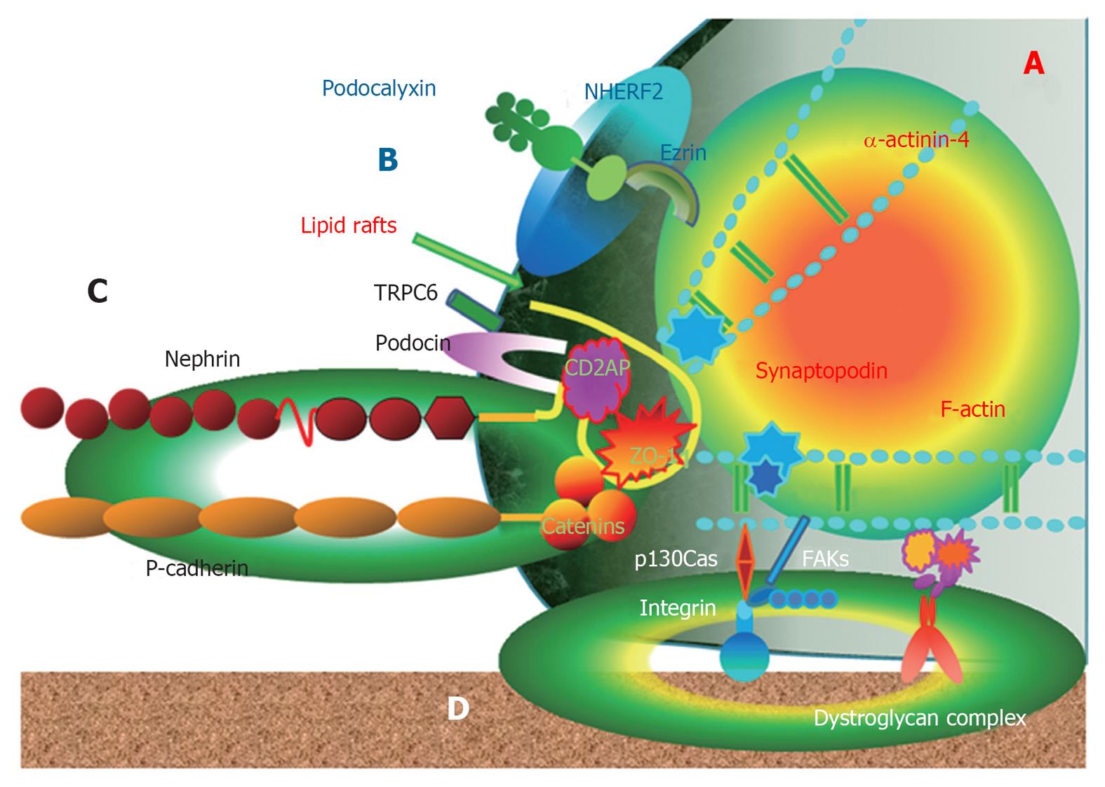
Figure 1 Schematic view of podocyte structure.
A: Cytoskeleton; B: Apical membrane domain; C: Slit diaphragm protein complex domain; D: Basal membrane domain. Adaptor proteins link mainly slit diaphragm proteins to cytoskeleton. FAKs: Focal adhesion kinases; ZO: Zonula occludens; CD2AP: CD2-associated protein.
- Citation: Ha TS. Roles of adaptor proteins in podocyte biology. World J Nephrol 2013; 2(1): 1-10
- URL: https://www.wjgnet.com/2220-6124/full/v2/i1/1.htm
- DOI: https://dx.doi.org/10.5527/wjn.v2.i1.1


