







Copyright: ©Author(s) 2026.
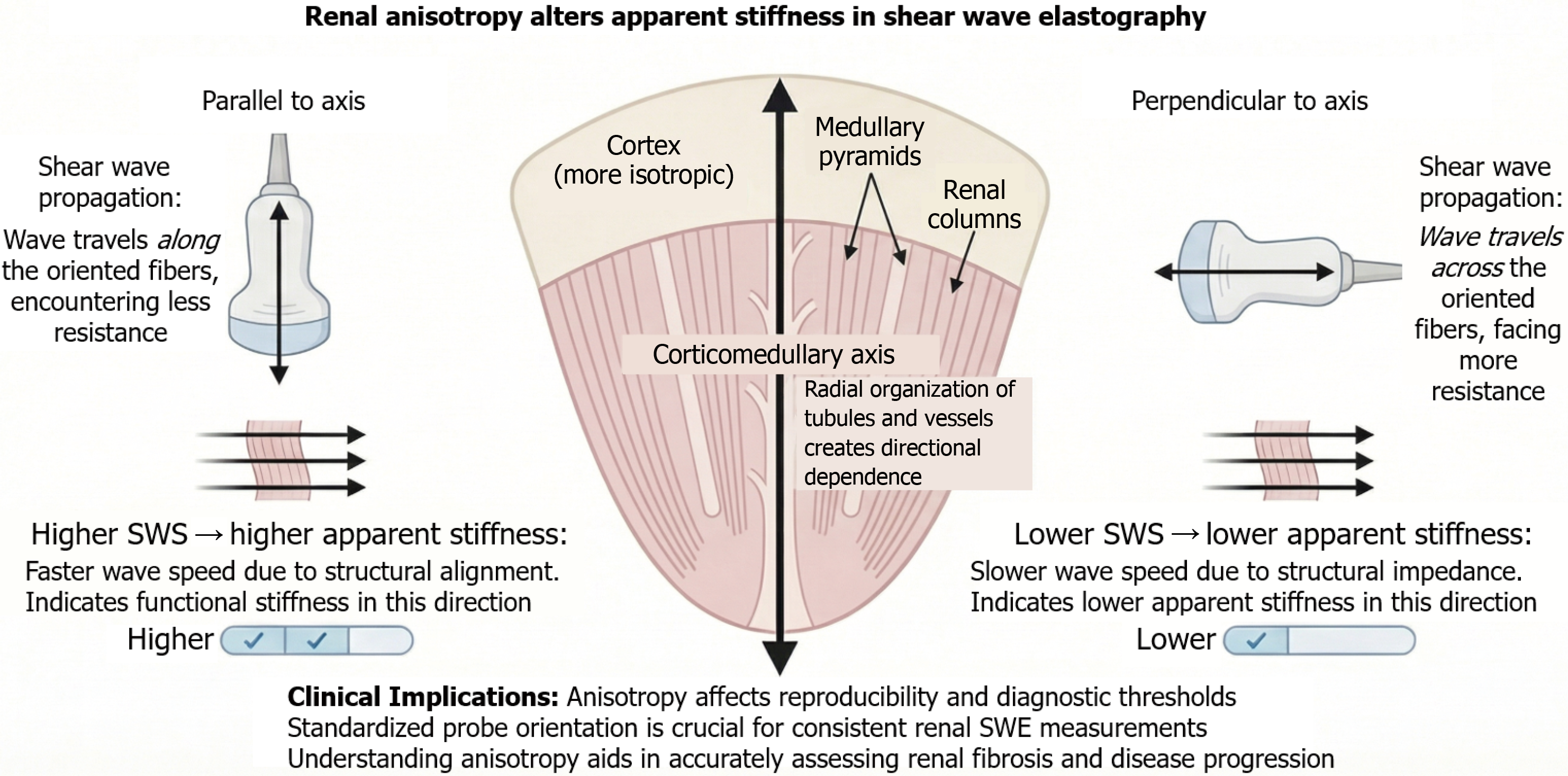
Figure 2 Effect of renal anisotropy on shear wave elastography measurements.
Renal tissue anisotropy significantly influences shear wave propa gation and, consequently, stiffness measurements obtained by shear wave elastography. When the ultrasound beam is aligned parallel to the corticomedullary axis, shear waves travel along tubular and vascular structures with less resistance, resulting in higher measured velocities and increased apparent stiffness. In contrast, perpendicular orientation results in slower wave propagation and lower stiffness due to higher structural impedance. These orientation-dependent differences highlight the impact of renal architecture on elastographic measurements and underscore the need for standardized probe positioning to improve reproducibility and interpretation in clinical practice. SWE: Shear-wave elastography; SWS: Shear-wave speed.
- Citation: Mora-Loján JL, García-Trujillo OF, Vélez-Hurtado G, Bermúdez-Mercado E, Pacheco-Salazar F, Balderas-Juarez J, Martínez-Sánchez FD. Renal elastography in kidney disease: Current evidence, limitations, and future directions. World J Nephrol 2026; 15(3): 119841
- URL: https://www.wjgnet.com/2220-6124/full/v15/i3/119841.htm
- DOI: https://dx.doi.org/10.5527/wjn.119841