
















Copyright: ©Author(s) 2026.
World J Nephrol. Jun 25, 2026; 15(2): 118343
Published online Jun 25, 2026. doi: 10.5527/wjn.v15.i2.118343
Published online Jun 25, 2026. doi: 10.5527/wjn.v15.i2.118343
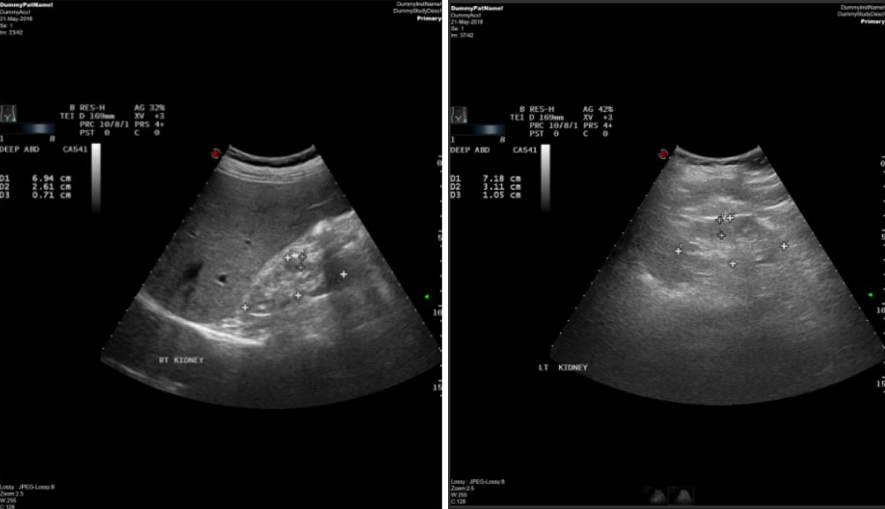
Figure 1 Ultrasound images of enlarged kidneys.
Ultrasound images of the kidneys of one patient on hemodialysis. Both kidneys are small with mildly to moderately increased parenchymal echogenicity and decreased parenchymal sinus differentiation. The right kidney measured 6.9 cm in length and 0.7 cm in parenchymal thickness, showing focal upper calyceal dilatation and irregularity.
- Citation: Almuhanna AA, Vatte C, Guo Q, Elsalamouni TS, Al-Muhanna FA, Aboalrihy AM, Alhabib HA, Almomen MF, Alali RA, Habara AH, Alrubaish MA, Alfalah KM, Cyrus C, Abdul-Rahman IS, Keating BJ, Al-Ali AK, Wang C. Gut microbiota in a Saudi population with chronic kidney disease. World J Nephrol 2026; 15(2): 118343
- URL: https://www.wjgnet.com/2220-6124/full/v15/i2/118343.htm
- DOI: https://dx.doi.org/10.5527/wjn.v15.i2.118343