
















Copyright: ©Author(s) 2026.
World J Nephrol. Jun 25, 2026; 15(2): 117336
Published online Jun 25, 2026. doi: 10.5527/wjn.v15.i2.117336
Published online Jun 25, 2026. doi: 10.5527/wjn.v15.i2.117336
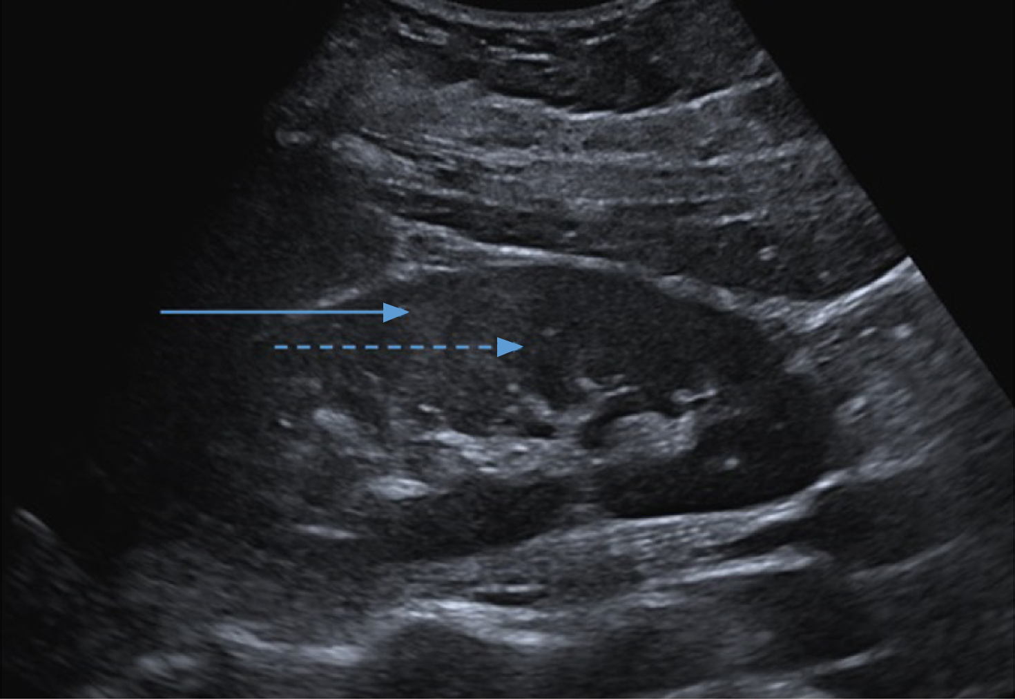
Figure 2 Ultrasound of the right kidney demonstrating preserved corticomedullary differentiation in which cortex is hyperechoic (solid line) relative to medulla (dashed line).
- Citation: Yaxley J, Scott T, Burnett C, Kurtkoti J. Performing a percutaneous kidney biopsy. World J Nephrol 2026; 15(2): 117336
- URL: https://www.wjgnet.com/2220-6124/full/v15/i2/117336.htm
- DOI: https://dx.doi.org/10.5527/wjn.v15.i2.117336