
















Copyright: ©Author(s) 2026.
World J Nephrol. Mar 25, 2026; 15(1): 114185
Published online Mar 25, 2026. doi: 10.5527/wjn.v15.i1.114185
Published online Mar 25, 2026. doi: 10.5527/wjn.v15.i1.114185
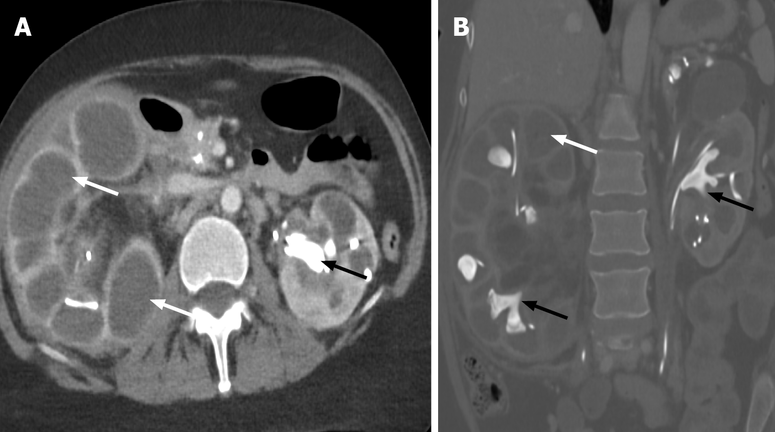
Figure 14 Xanthogranulomatous pyelonephritis.
A and B: Axial (A) and coronal (B) computed tomography demonstrating diffusely dilated renal calyceal system with renal cortical thinning (bear paw sign) (white arrow). Multiple staghorn calculi are filling the calyces (indicated by the black arrow). These findings are consistent with xanthogranulomatous pyelonephritis.
- Citation: Vulasala SSR, Robinson J, Engel C, Zulia Y, Sharma A, Gopireddy DR, Adler G, Virarkar M. Computed tomography of renal emergencies: A comprehensive diagnostic guide for radiology residents. World J Nephrol 2026; 15(1): 114185
- URL: https://www.wjgnet.com/2220-6124/full/v15/i1/114185.htm
- DOI: https://dx.doi.org/10.5527/wjn.v15.i1.114185