
















Copyright: ©Author(s) 2026.
World J Nephrol. Mar 25, 2026; 15(1): 114185
Published online Mar 25, 2026. doi: 10.5527/wjn.v15.i1.114185
Published online Mar 25, 2026. doi: 10.5527/wjn.v15.i1.114185
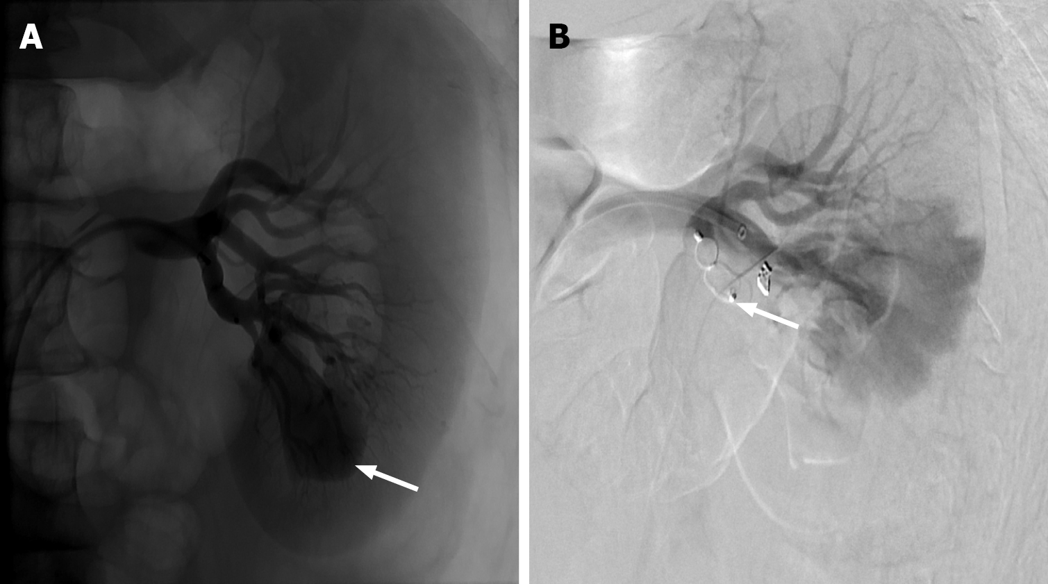
Figure 9 Renal arterio-venous fistula.
A: Left renal artery angiogram demonstrating a large inferior pole pseudoaneurysm (white arrow); B: Subsequent treatment with an Amplatzer plug (white arrow) and a single coil, with successful occlusion of the pseudoaneurysm. Additionally, early venous filling was noted during examination, concerning for traumatic renal arterio-venous fistula.
- Citation: Vulasala SSR, Robinson J, Engel C, Zulia Y, Sharma A, Gopireddy DR, Adler G, Virarkar M. Computed tomography of renal emergencies: A comprehensive diagnostic guide for radiology residents. World J Nephrol 2026; 15(1): 114185
- URL: https://www.wjgnet.com/2220-6124/full/v15/i1/114185.htm
- DOI: https://dx.doi.org/10.5527/wjn.v15.i1.114185