
















Copyright: ©Author(s) 2026.
World J Nephrol. Mar 25, 2026; 15(1): 114185
Published online Mar 25, 2026. doi: 10.5527/wjn.v15.i1.114185
Published online Mar 25, 2026. doi: 10.5527/wjn.v15.i1.114185
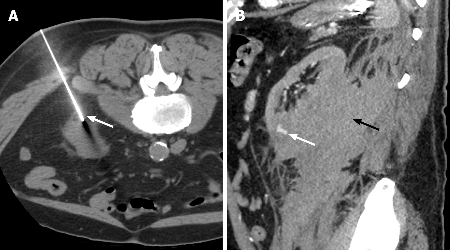
Figure 3 Grade III renal Injury.
A: Non-contrast computed tomography obtained during biopsy of the left kidney with the patient in the prone position, demonstrating the biopsy needle with the tip abutting the left inferior renal pole (white arrow); B: Follow-up arterial phase computed tomography abdomen 48 hours later demonstrates left subcapsular and retroperitoneal haemorrhage (black arrow) with an area of contrast blush in the inferior renal pole consistent with pseudoaneurysm (white arrow).
- Citation: Vulasala SSR, Robinson J, Engel C, Zulia Y, Sharma A, Gopireddy DR, Adler G, Virarkar M. Computed tomography of renal emergencies: A comprehensive diagnostic guide for radiology residents. World J Nephrol 2026; 15(1): 114185
- URL: https://www.wjgnet.com/2220-6124/full/v15/i1/114185.htm
- DOI: https://dx.doi.org/10.5527/wjn.v15.i1.114185