
















©The Author(s) 2025.
World J Nephrol. Dec 25, 2025; 14(4): 112066
Published online Dec 25, 2025. doi: 10.5527/wjn.v14.i4.112066
Published online Dec 25, 2025. doi: 10.5527/wjn.v14.i4.112066
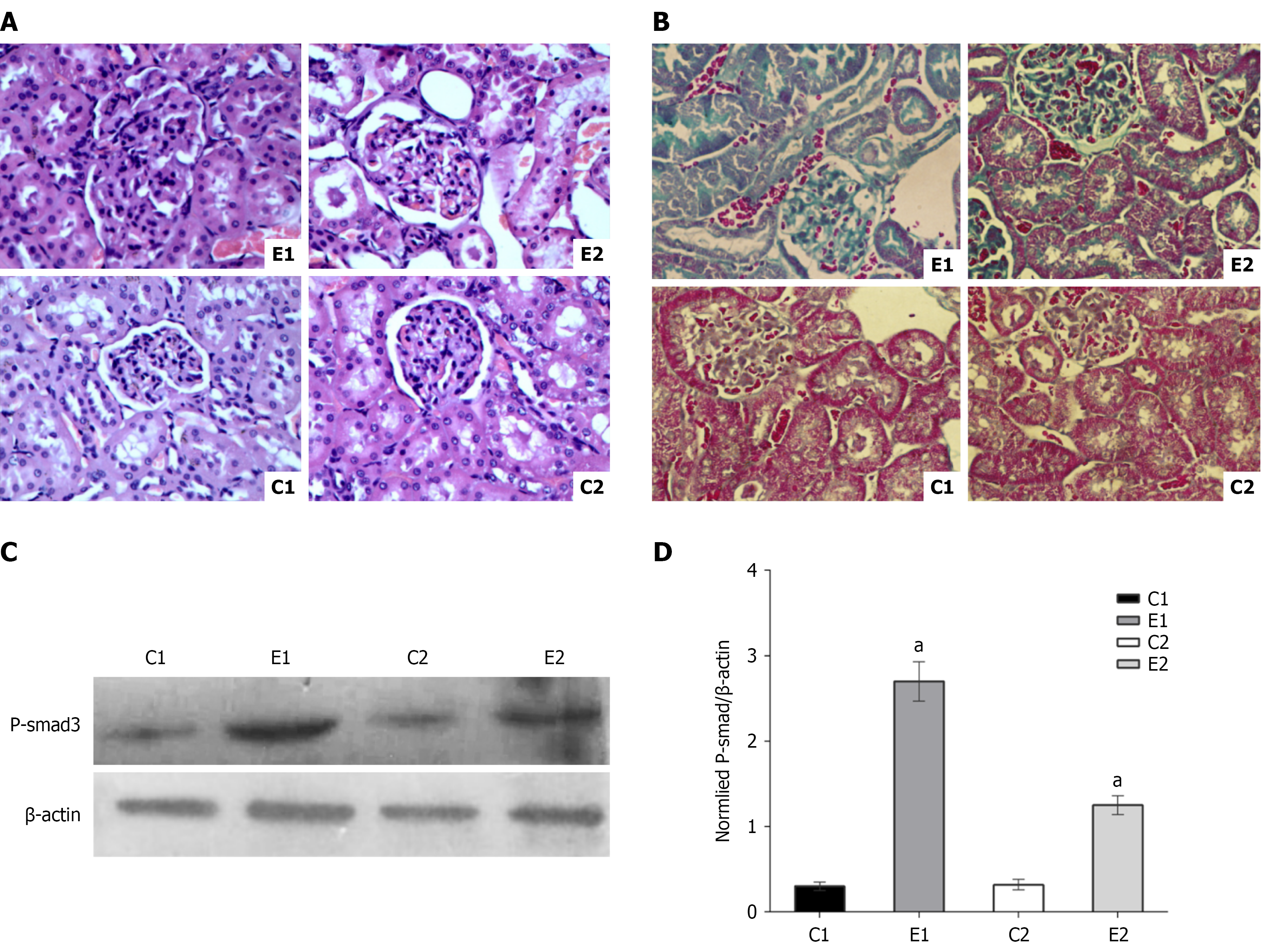
Figure 1 Effect of trimethylamine N-oxide on renal function injury.
aP < 0.01. A: Hematoxylin and eosin staining of rat kidney tissue (400 ×). Group E1 showed the most obvious structural damage to the kidneys, with proliferation of glomerular mesangial cells and mesangial matrix, and shrinkage of Bowman’s capsule. Renal tubular protein casts were also observed in the renal tubules. Group E2 showed milder damage to the renal tissue, while groups C1 and C2 showed normal renal structure; B: Masson staining of rat kidney to assess fibrosis (400 ×). Group E1: Diabetic nephropathy model group, group E2 diabetic model inhibition group, group C1 blank control group, group C2 blank inhibition group; fibrotic tissue is stained green in the figure; C: Level of the P-smad3 protein in different groups of rats. E1 group protein expression was significantly higher than other groups (P < 0.01), while E2 group protein expression was higher than C1 and C2 groups, but significantly lower than E1 group (P < 0.01); D: Gray values indicating the level of the P-smad3 protein in different groups of rats.
- Citation: Song YJ, Yang B, Feng QS, Ma FF, Xing B, Bin XL, Ha XQ. Gut microbiota-derived trimethylamine N-oxide exacerbates diabetic nephropathy by promoting renal fibrosis. World J Nephrol 2025; 14(4): 112066
- URL: https://www.wjgnet.com/2220-6124/full/v14/i4/112066.htm
- DOI: https://dx.doi.org/10.5527/wjn.v14.i4.112066