
















©The Author(s) 2025.
World J Nephrol. Dec 25, 2025; 14(4): 111613
Published online Dec 25, 2025. doi: 10.5527/wjn.v14.i4.111613
Published online Dec 25, 2025. doi: 10.5527/wjn.v14.i4.111613
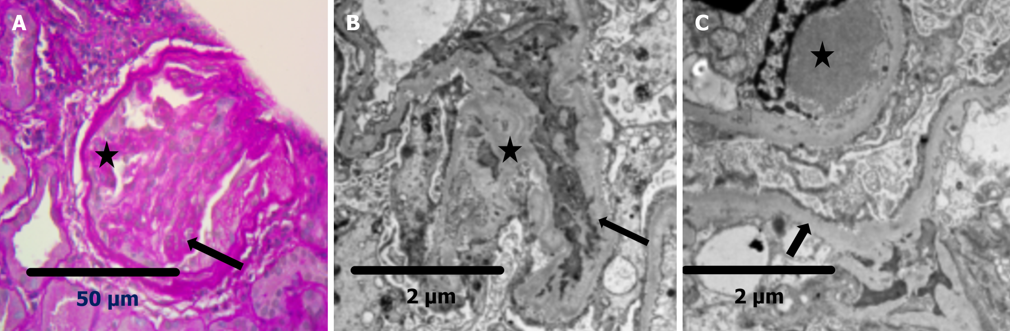
Figure 2 Shows focal segmental sclerosis with early diffuse diabetic nephropathy changes.
A: Segmental glomerular sclerosis showing obliteration of glomerular capillary lumina (arrow) and adhesion of the sclerotic tuft to Bowman’s capsule (star) (Periodic acid–Schiff stain, × 40); B: Ultrastructurally, the mesangium is expanded by increased mesangial matrix (star), and the glomerular basement membrane is thickened (arrow) (Transmission electron microscopy, uranyl acetate and lead citrate stain); C: Electron micrograph demonstrating loss of normal glomerular basement membrane ultrastructural detail (arrow) and subendothelial deposition of dense material consistent with hyalinosis (star) (Transmission electron microscopy, uranyl acetate and lead citrate stain).
- Citation: Al-Qurashi SH, Khalil MAM, Mahmood HHK, A Al-Ghamdi R, Alsharif MM, Said Ahmed MA, Alghamdi RMH, Sadagah NM. Predictors of non-diabetic kidney disease in diabetics: A Saudi Arabian perspective. World J Nephrol 2025; 14(4): 111613
- URL: https://www.wjgnet.com/2220-6124/full/v14/i4/111613.htm
- DOI: https://dx.doi.org/10.5527/wjn.v14.i4.111613