
















©The Author(s) 2025.
World J Nephrol. Dec 25, 2025; 14(4): 110491
Published online Dec 25, 2025. doi: 10.5527/wjn.v14.i4.110491
Published online Dec 25, 2025. doi: 10.5527/wjn.v14.i4.110491
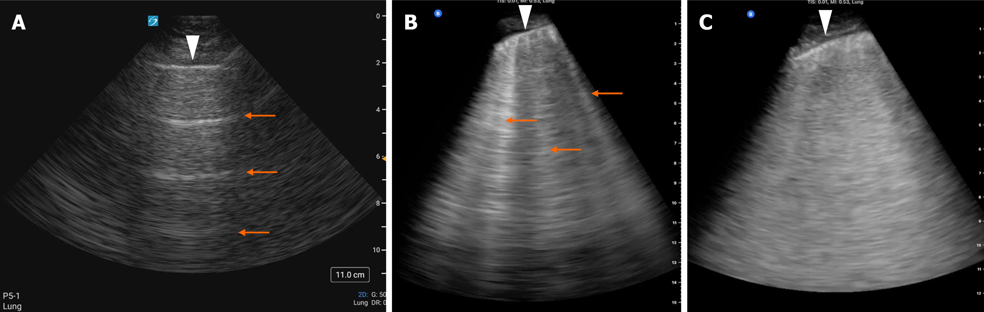
Figure 2 Lung ultrasound images showing.
A: Normal horizontal artifacts known as A-lines (indicated by arrows); B: Discrete B-lines (arrows); C: Confluent B-lines creating a white-out appearance in the scanned lung zone typically seen in alveolar edema. The arrowhead in each image indicates the pleural line.
- Citation: Gogula NSAR, Koratala A. Lung ultrasound in nephrology: Basics, applications, limitations, and future directions. World J Nephrol 2025; 14(4): 110491
- URL: https://www.wjgnet.com/2220-6124/full/v14/i4/110491.htm
- DOI: https://dx.doi.org/10.5527/wjn.v14.i4.110491