
















©The Author(s) 2025.
World J Nephrol. Sep 25, 2025; 14(3): 108703
Published online Sep 25, 2025. doi: 10.5527/wjn.v14.i3.108703
Published online Sep 25, 2025. doi: 10.5527/wjn.v14.i3.108703
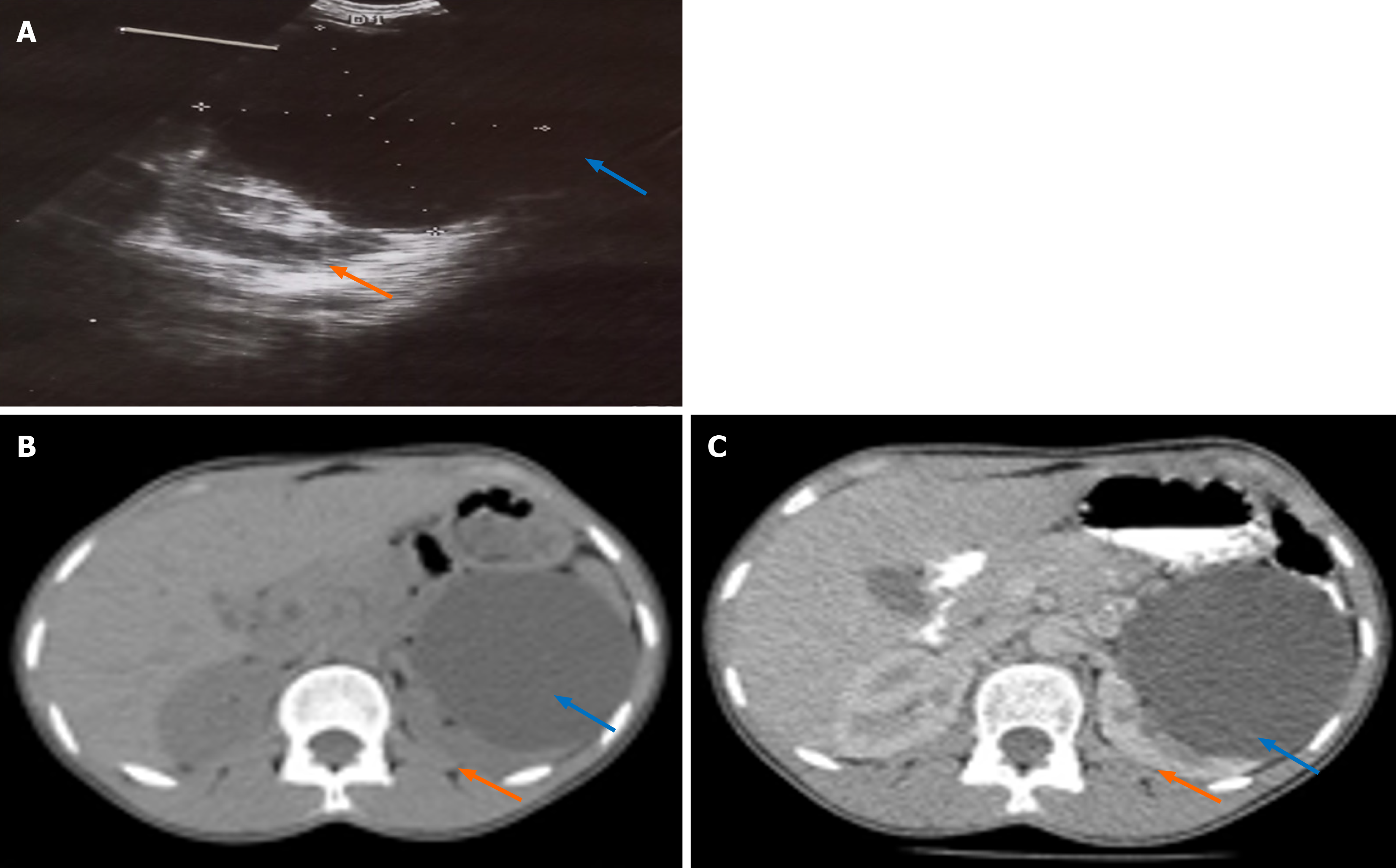
Figure 1 Imaging studies.
A: Transabdominal ultrasound scan films demonstrating the left renal cyst and compressed left kidney (blue and orange arrows) respectively; B and C: Non-contrast and contrast-enhanced abdominal computerized tomography scan of the same pathology with bilaterally functioning kidneys.
- Citation: Khalid A, Yakubu KB, Umar AM, Aljannare BG, Aminu NA, Obadele OG, Abdulwahab-Ahmed A. Uncommon presentation and management of a giant renal cyst abscess: A case report. World J Nephrol 2025; 14(3): 108703
- URL: https://www.wjgnet.com/2220-6124/full/v14/i3/108703.htm
- DOI: https://dx.doi.org/10.5527/wjn.v14.i3.108703