
















©The Author(s) 2025.
World J Nephrol. Sep 25, 2025; 14(3): 108534
Published online Sep 25, 2025. doi: 10.5527/wjn.v14.i3.108534
Published online Sep 25, 2025. doi: 10.5527/wjn.v14.i3.108534
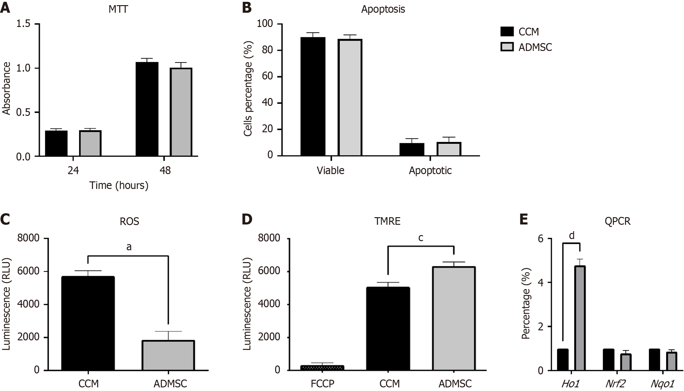
Figure 2 Effects of adipose-derived mesenchymal stromal cells treatment compared to cell culture media on cellular function and oxidative stress.
A: MTT assay showing cellular metabolic activity at 24 hours and 48 hours. Absorbance values indicate no significant difference between adipose-derived mesenchymal stromal cells (ADMSCs) and cell culture media (CCM) groups; B: Apoptosis analysis by flow cytometry indicating similar percentages of viable and apoptotic cells between groups; C: Intracellular reactive oxygen species (ROS) levels measured by luminescence. ADMSC treatment significantly reduced ROS levels compared to CCM (aP < 0.05); D: Mitochondrial membrane potential assessed by tetramethylrhodamine ethyl ester (TMRE) staining. ADMSC-treated cells showed significantly higher TMRE fluorescence, indicating improved mitochondrial function (cP < 0.001); E: Heme oxygenase 1 (HO-1), nuclear factor erythroid 2-related factor 2 (Nrf2), and NAD(P)H quinone dehydrogenase 1 (Nqo1) mRNA levels were measured using quantitative PCR. Relative expression of HO-1, Nrf2, and Nqo1 was calculated and normalized to the b-actin housekeeping gene control. Each value represents the mean ± SD (n = 4) (dP < 0.0001). BM-MSCs: Bone marrow-derived mesenchymal stromal cells.
- Citation: Jafar H, Ababneh NA, Alhattab D, Alatoom RM, Zalloum S, Salah B, Alhawari H, Awidi A. Adipose-derived mesenchymal stromal cell secretome protects against kidney injury through induction of heme oxygenase 1 upregulation in vitro. World J Nephrol 2025; 14(3): 108534
- URL: https://www.wjgnet.com/2220-6124/full/v14/i3/108534.htm
- DOI: https://dx.doi.org/10.5527/wjn.v14.i3.108534