
















©The Author(s) 2025.
World J Nephrol. Sep 25, 2025; 14(3): 107571
Published online Sep 25, 2025. doi: 10.5527/wjn.v14.i3.107571
Published online Sep 25, 2025. doi: 10.5527/wjn.v14.i3.107571
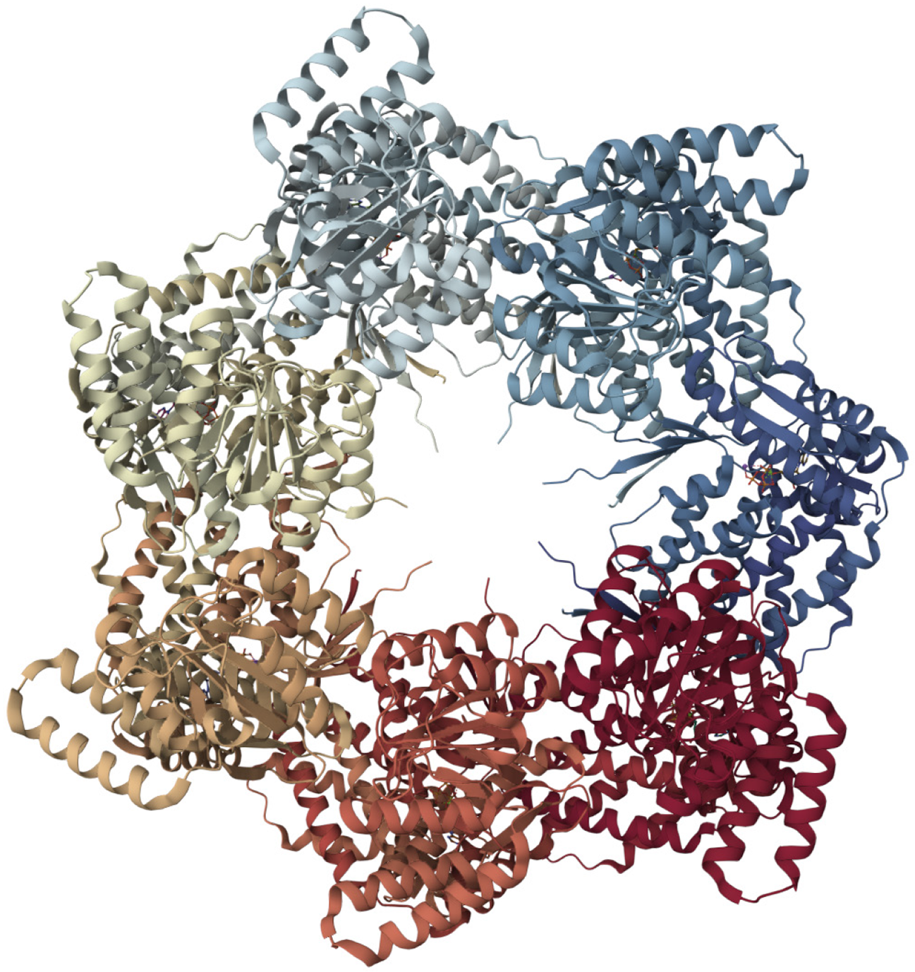
Figure 2 Molecular structure of heat shock protein 60[82].
Each heat shock protein 60 (HSP60) monomer features three key domains: The apical domain for binding substrates and the co-chaperone HSP10, an intermediate domain acting as a hinge, and an equatorial domain responsible for adenosine triphosphate (ATP) binding and inter-ring contacts. Notably, upon ATP binding, mitochondrial HSP60's apical domains adopt an asymmetric "up/down" configuration, a crucial feature for simultaneously recruiting HSP10 and engaging client proteins, thereby coordinating the folding process. This entire ATP-dependent mechanism involves significant conformational shifts that regulate the folding chamber, enabling HSP60 to efficiently fold proteins within the mitochondria. This figure was adapted with Mol* (MIT-licensed) from the Structure page for PDB ID 8G7M (CC0 1.0) on the RCSB Protein Data Bank (https://www.rcsb.org/structure/8G7M). Citation: Braxton JR, Shao H, Tse E, Gestwicki JE, Southworth DR. Asymmetric apical domain states of mitochondrial Hsp60 coordinate substrate engagement and chaperonin assembly. Nat Struct Mol Biol 2024; 31: 1848-1858. Copyright ©The Author(s) 2024. Published by Nature Pub. Group (Supplementary material).
- Citation: Tran TTT, Tran KV, Nguyen TD, Pham NTT, Nguyen TH. Role of heat shock proteins in renal function and adaptation to heat stress: Implications for global warming. World J Nephrol 2025; 14(3): 107571
- URL: https://www.wjgnet.com/2220-6124/full/v14/i3/107571.htm
- DOI: https://dx.doi.org/10.5527/wjn.v14.i3.107571