Copyright
©The Author(s) 2017.
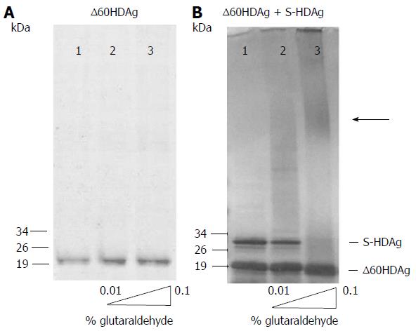
Figure 2 S-HDAg and ∆60HDAg multimerization ability.
In panels A and B, purified recombinant protein was cross-linked with increasing concentrations of glutaraldehyde (0%, 0.01% and 0.1%) prior to SDS-PAGE. Proteins were detected by Coomassie blue staining. In panel A, only purified ∆60HDAg was present at 2 μmol/L and in panel B both S-HDAg and ∆60HDAg were present at 2 μmol/L each. The arrow indicates the presence high molecular weight oligomers.
- Citation: Alves C, Cheng H, Tavanez JP, Casaca A, Gudima S, Roder H, Cunha C. Structural and nucleic acid binding properties of hepatitis delta virus small antigen. World J Virol 2017; 6(2): 26-35
- URL: https://www.wjgnet.com/2220-3249/full/v6/i2/26.htm
- DOI: https://dx.doi.org/10.5501/wjv.v6.i2.26


