
















©The Author(s) 2025.
World J Virol. Dec 25, 2025; 14(4): 115071
Published online Dec 25, 2025. doi: 10.5501/wjv.v14.i4.115071
Published online Dec 25, 2025. doi: 10.5501/wjv.v14.i4.115071
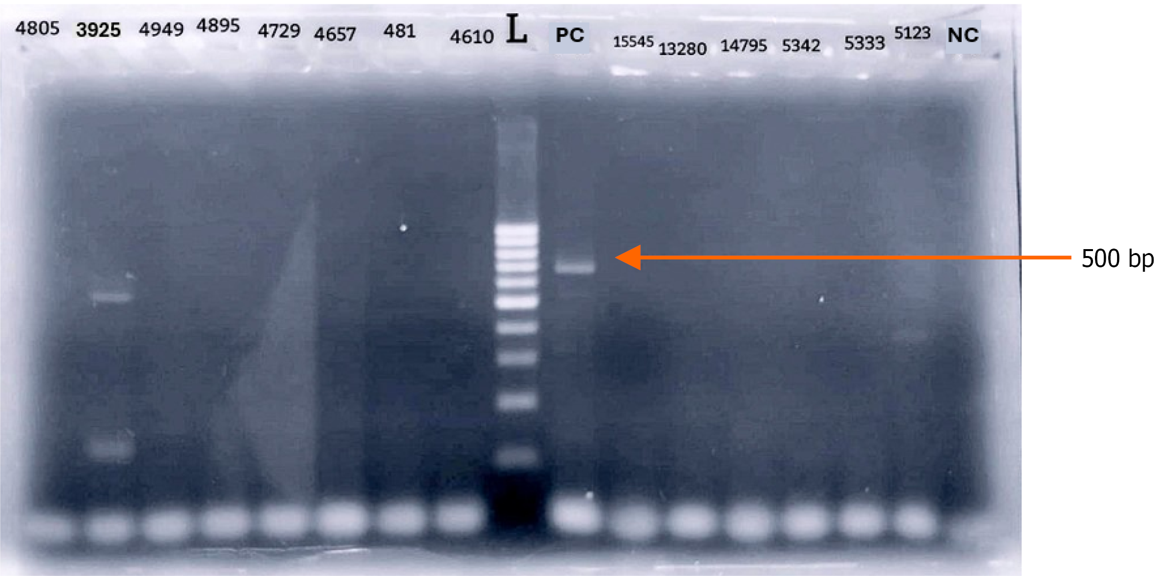
Figure 2 Gel electrophoresis image showing reverse transcriptase polymerase chain reaction amplification of cerebrospinal fluid samples using flavivirus-universal primers.
The amplified products were visualized with Ethidium bromide staining. L: 100 bp DNA ladder; PC: Positive control (Japanese encephalitis virus); NC: Negative control; Sample ID 3925: A patient sample showing a distinct band between 500-600 bp; Other lanes: Non-reactive cerebrospinal fluid samples. Arrow: Positive control in par with 500 pb of the ladder.
- Citation: Perera I, Weerathunga A, Arachchige N, Rajamanthri L, Fernando S, Muthugala R. Detection of dengue virus encephalitis in central part of Sri Lanka. World J Virol 2025; 14(4): 115071
- URL: https://www.wjgnet.com/2220-3249/full/v14/i4/115071.htm
- DOI: https://dx.doi.org/10.5501/wjv.v14.i4.115071