Copyright
©2012 Baishideng Publishing Group Co.
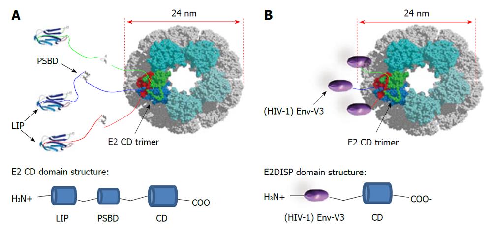
Figure 2 Schematic image of the E2 acetyltransferase display system derived from the Bacillus stearothermophilus pyruvate dehydrogenase complex.
A: The E2 chain as it occurs in the native pyruvate dehydrogenase complex. Twenty trimers of the E2 polypeptide chain form a pentagonal dodecahedron (60-mer) with icosahedral symmetry; B: Representation of the E2 core displaying the HIV-1 Envelope (Env) third hypervariable region V3 on the surface of the scaffold. LIP: Lypoil domain; PSBD: Peripheral subunit-binding domain; CD: Catalytic domain; E2DISP: E2 acetyltransferase display system.
- Citation: Trovato M, Krebs SJ, Haigwood NL, De Berardinis P. Delivery strategies for novel vaccine formulations. World J Virol 2012; 1(1): 4-10
- URL: https://www.wjgnet.com/2220-3249/full/v1/i1/4.htm
- DOI: https://dx.doi.org/10.5501/wjv.v1.i1.4


