Copyright
©The Author(s) 2016.
World J Virol. Aug 12, 2016; 5(3): 125-134
Published online Aug 12, 2016. doi: 10.5501/wjv.v5.i3.125
Published online Aug 12, 2016. doi: 10.5501/wjv.v5.i3.125
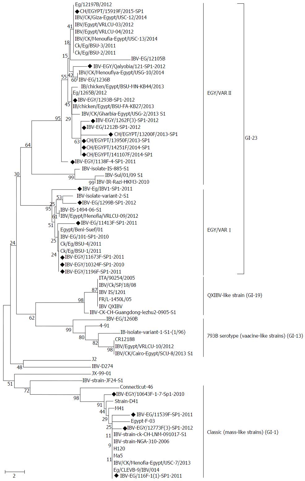
Figure 1 Phylogenetic tree representing the partial amino acid sequences of the S1 gene for 20 infectious bronchitis virus isolates (marked with black diamond) with other related infectious bronchitis virus and reference strains.
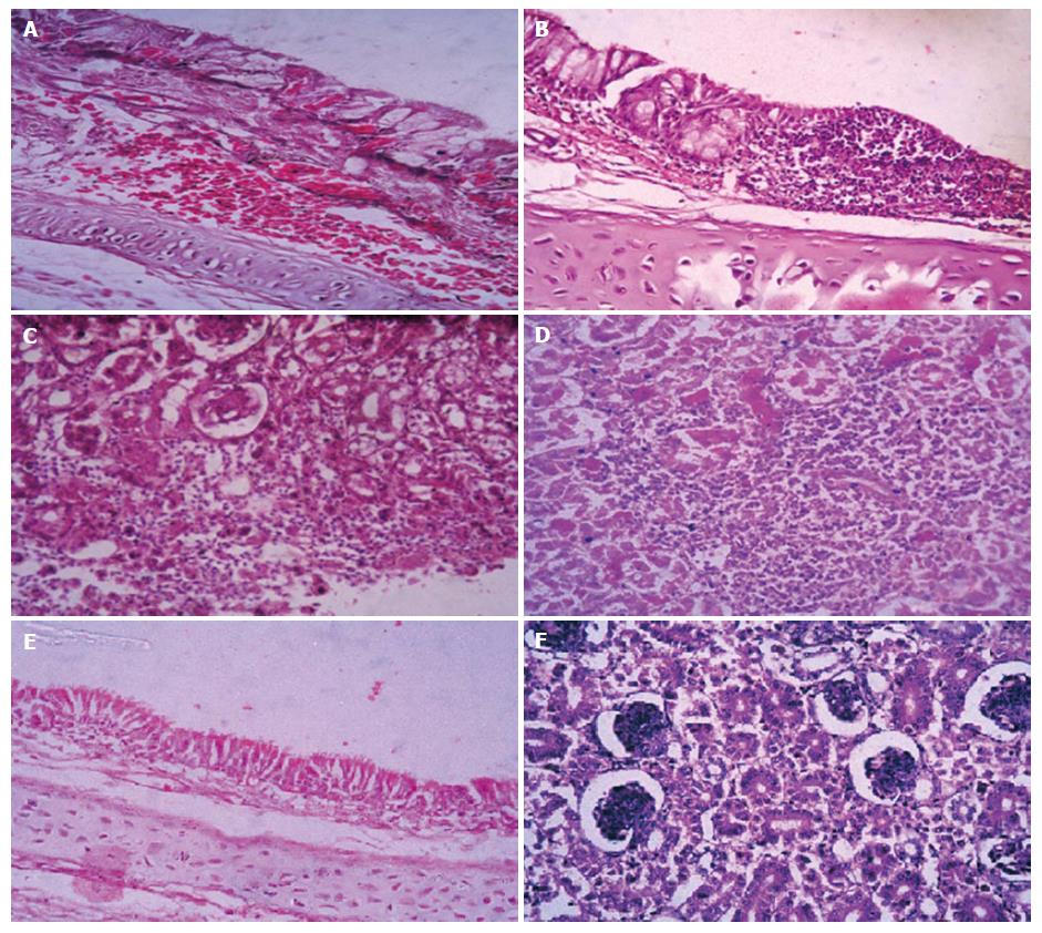
Figure 2 Histopathology illustration of the trachea and kidney from experimentally infected chickens.
A: Trachea (from Group 1) showed subepithelial hemorrhage accompanied with goblet cell activation, inflammatory cells and edema; B: Trachea (from Group 2) with focal aggregation of lymphocytic cells and epithelial desquamation, ulceration accompanied with goblet cell hypertrophy; C: Kidney (from Group 2) showed glomerular edema and glomerulonephritis with extravasation of blood vessels between renal tubules; D: Kidney (from Group 1) with severe necrosis of renal tubules and focal lymphocytic aggregation; E: Trachea of negative control group; F: Kidney of negative control group (H and E, × 20).
- Citation: Zanaty A, Arafa AS, Hagag N, El-Kady M. Genotyping and pathotyping of diversified strains of infectious bronchitis viruses circulating in Egypt. World J Virol 2016; 5(3): 125-134
- URL: https://www.wjgnet.com/2220-3249/full/v5/i3/125.htm
- DOI: https://dx.doi.org/10.5501/wjv.v5.i3.125


