Copyright
©The Author(s) 2016.
World J Transplant. Jun 24, 2016; 6(2): 278-290
Published online Jun 24, 2016. doi: 10.5500/wjt.v6.i2.278
Published online Jun 24, 2016. doi: 10.5500/wjt.v6.i2.278
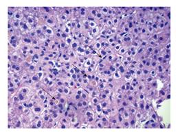
Figure 2 Progressive familial intrahepatic cholestasis type 1 with severe bland lobular cholestasis and lobular disarray.
The image shows bile plugging with surrounding pseudorosette formation (arrows). In PFIC1, the canalicular bile is course on electronic microscopy and also referred to as “Byler bile”. Thick bile is seen within the pseudorosette here on H and E stain. There is an absence of lobular inflammation and typically no features of neonatal giant cell hepatitis. PFIC: Progressive familial intrahepatic cholestasis.
- Citation: Mehl A, Bohorquez H, Serrano MS, Galliano G, Reichman TW. Liver transplantation and the management of progressive familial intrahepatic cholestasis in children. World J Transplant 2016; 6(2): 278-290
- URL: https://www.wjgnet.com/2220-3230/full/v6/i2/278.htm
- DOI: https://dx.doi.org/10.5500/wjt.v6.i2.278


