Copyright
©2013 Baishideng Publishing Group Co.
World J Transplant. Jun 24, 2013; 3(2): 26-29
Published online Jun 24, 2013. doi: 10.5500/wjt.v3.i2.26
Published online Jun 24, 2013. doi: 10.5500/wjt.v3.i2.26
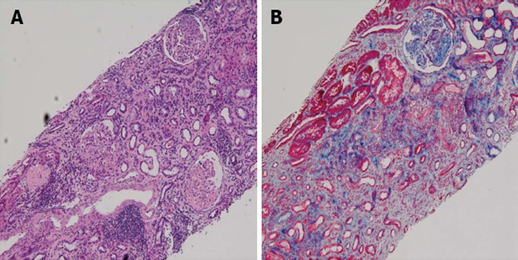
Figure 1 Patient was admitted to the hospital and underwent a diagnostic percutaneous ultrasound guided renal biopsy (HE stain, ×100).
Hematoxylin and eosin (A) and periodic acid-Schiff stains of the kidney biopsy specimens (light microscopy) (B) showing the histopathology examination of the kidney, which tissue confirm the presence of focal segmental glomerulosclerosis as evidenced by involvement of approximately 50% of the glomeruli with segmental lesions and some of the glomeruli had total global glomerulosclerosis. There was also associated interstitial fibrosis and tubular atrophy.
- Citation: Elmahi N, Csongrádi &, Kokko K, Lewin JR, Davison J, Fülöp T. Residual renal function in peritoneal dialysis with failed allograft and minimum immunosuppression. World J Transplant 2013; 3(2): 26-29
- URL: https://www.wjgnet.com/2220-3230/full/v3/i2/26.htm
- DOI: https://dx.doi.org/10.5500/wjt.v3.i2.26


