
















Copyright: ©Author(s) 2026.
World J Transplant. Sep 18, 2026; 16(3): 120920
Published online Sep 18, 2026. doi: 10.5500/wjt.120920
Published online Sep 18, 2026. doi: 10.5500/wjt.120920
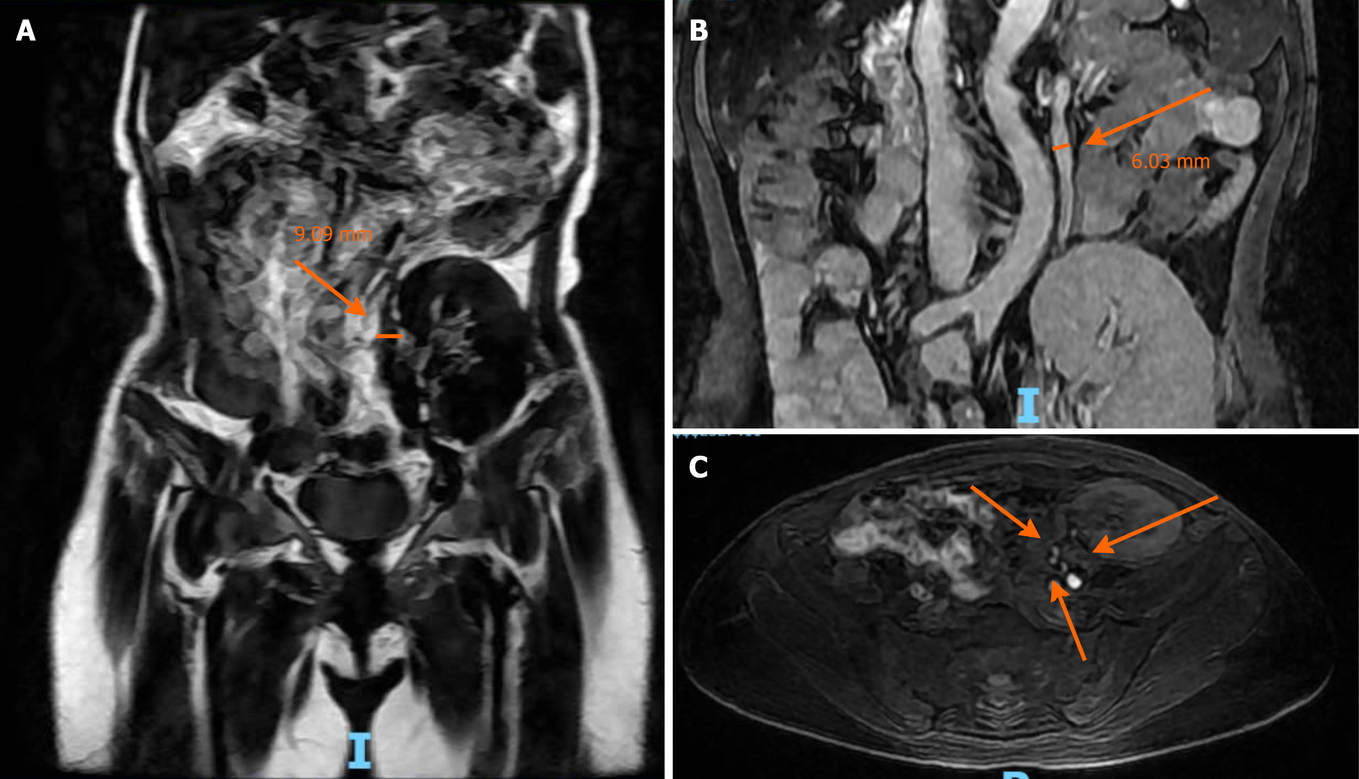
Figure 4 Follow-up magnetic resonance imaging at 8 months.
A: Great saphenous vein conduit dilated to 9.09 mm (arrow); B: Left ovarian vein enlarged to 6.03 mm (arrow); C: Patent triple arterial anastomoses supplying the renal graft (arrows).
- Citation: Nguyen DD, Pham HN, Nguyen HK, Bui KCL, Vo TM, Pham HN. Ovarian vein bypass for renal allograft salvage in iliac vein atrophy: A case report and review of literature. World J Transplant 2026; 16(3): 120920
- URL: https://www.wjgnet.com/2220-3230/full/v16/i3/120920.htm
- DOI: https://dx.doi.org/10.5500/wjt.120920