
















©The Author(s) 2026.
World J Transplant. Mar 18, 2026; 16(1): 114162
Published online Mar 18, 2026. doi: 10.5500/wjt.v16.i1.114162
Published online Mar 18, 2026. doi: 10.5500/wjt.v16.i1.114162
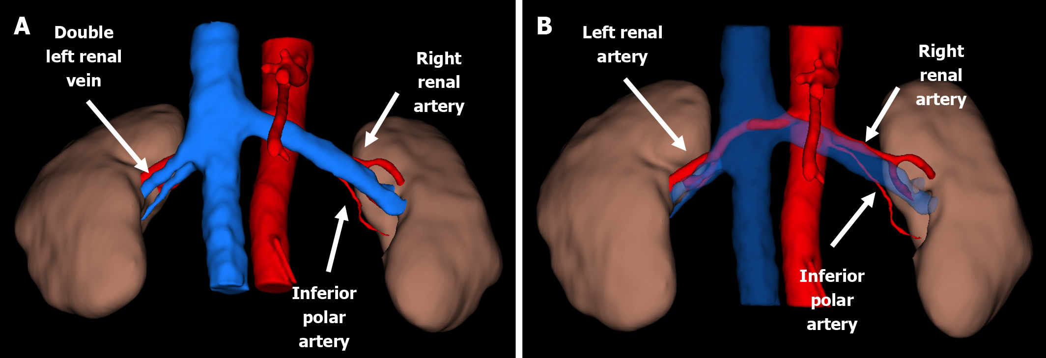
Figure 2 Three-dimensional reconstruction of computed tomography angiography illustrating the anatomical configuration of the donor renal arteries.
A: Anterior view; B: Anterior view with the left renal vein digitally removed to highlight the left renal arteries.
- Citation: Lekehal B, Ait Youssef N, Lekehal M, Jdar A, El Hassani AEA, Belyazid I, Bakkali T, Bounssir A. Acute graft thrombosis in a patient with factor V Leiden mutation: A case report and review of literature. World J Transplant 2026; 16(1): 114162
- URL: https://www.wjgnet.com/2220-3230/full/v16/i1/114162.htm
- DOI: https://dx.doi.org/10.5500/wjt.v16.i1.114162